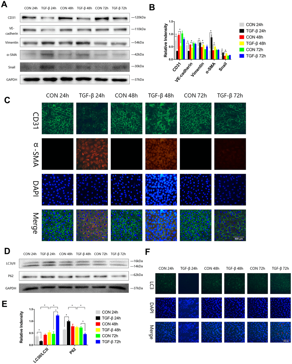
Figure 1.The autophagy level was changed during TGF-β-induced EndMT. (A, B) HUVECs were exposed to TGF-β (20 ng/ml) for 24, 48 and 72 h. Endothelial and mesenchymal markers were assessed using antibodies against CD31, VE-cadherin, Vimentin, α-SMA and Snail via Western blot. (C) Representative images (Scale bars= 500 μm) of immunofluorescence after staining of CD31 and α-SMA 24, 48 and 72 h after TGF-β stimulation. (D, E) Immunoblots were probed for autophagy markers LC3-II/LC3-I and p62 of cell lysates harvested from HUVECs treated with TGF-β for 24, 48 and 72 h. (F) Representative fluorescence microphotographs (Scale bars= 500 μm) of HUVECs immunostained for LC3. Bar graphs represent data that were from three independent experiments, and data represent the means±SEM. Unpaired T test (*P<0.05) was used to compare the significances between two groups.