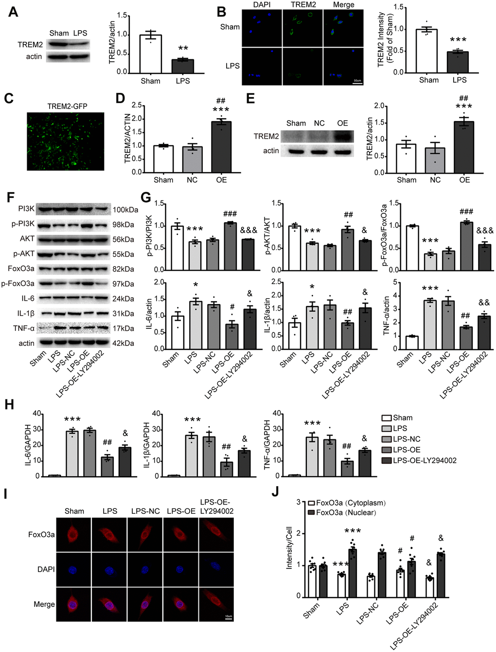
Figure 6.TREM2 overexpression activates the PI3K/AKT/FoxO3a pathway and inhibits the inflammatory response in BV2 cells. (A) Meaningful western blot bands and gray analysis of TREM2 (n=3), **P<0.01 vs Sham. (B) Confocal images and quantification analysis of fluorescence intensity of TREM2 (n=5). Scale bar, 50 μm. ***P<0.001 vs Sham. (C) A image of the overexpression of TREM2 plasmid after cell transfection. (D, E) TRME2 mRNA and protein were detected by qPCR and Western blot, respectively, in whole-cell lysates (n=4). ***P<0.001 vs Sham; ## P<0.01 vs NC. (F, G) Representative western blot bands (F) and its quantification (G) of p-PI3K, p-AKT, p-FoxO3, IL-6, IL-1β and TNF-α (n=4). (H) mRNA levels of proinflammatory cytokines (IL-1β, IL-6, TNF-α) were examined by qPCR (n=4). (I) The FoxO3a (red) were stained in the cells and observed by confocal microscope. (J) The quantification of FoxO3a in both nucleus and cytoplasm in each group (n=8); Magnification= 40x. Scale bar=10μm. * P<0.05, ** P<0.01, *** P<0.001, vs Sham; # P<0.05, ## P<0.01, ### P<0.001, vs LPS-NC; & P<0.05, && P<0.01, &&& P<0.001, vs LPS-OE. Results were represented as mean ± SEM.