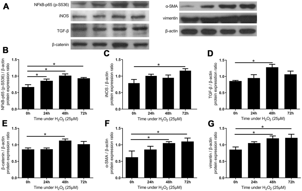
Figure 5.The expressions of inflammatory markers and fibrotic markers increased after administration of H2O2-induced oxidative stress. (A) The expression of inflammatory markers and fibrotic markers. The quantified protein expression ratio of (B) phosphorylated NF-κB p65 (S536) and (C) iNOS increased substantially under oxidative stress, suggesting the activation of inflammatory processes. The expressions of fibrotic signals such as (D) TGF-β and (E) β-catenin increased after 48 hours, followed by increases in fibrotic markers including (F) α-SMA and (G) vimentin. (n = 6; * p < 0.05 compared to the control group (0’); the value was generated by one-way ANOVA and post-hoc Tukey’s test.).