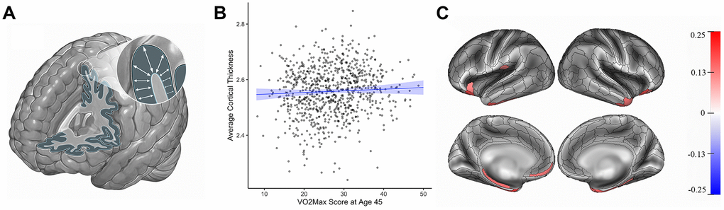
Figure 1.Cortical thickness (mm) and cardiovascular fitness (mL/min/kg) at age 45. (A) Cortical thickness. (B) Graph showing the correlation between average cortical thickness (mm, y-axis) and VO2Max (mL/min/kg; x-axis). Average cortical thickness was unrelated to the VO2Max scores of Study members (β=0.05, 95% CI = -0.04 to 0.14 p=0.28). (C) Study members with higher VO2Max scores, however, had increased parcel-wise thickness in multiple regions encompassing anterior temporal cortex, parahippocampal gyrus, and prefrontal cortex. Color bar on the right of the figure indicates a possible range of βs from -.25 to +.25. The color of each parcel on the simulated brains represents the associated effect size with cardiovascular fitness. Parcels colored in gray did not remain significantly associated with cardiovascular fitness after adjusting for multiple comparisons. All results pictured are adjusted for sex. mL/min/kg = milliliters per minute per kilogram; VO2Max = volume of maximum oxygen uptake; β = standardized coefficient; CI = confidence interval.