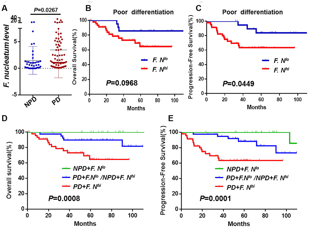
Figure 3.The association between F. nucleatum levels and tumor histological differentiation. (A) F. nucleatum burdens in 112 tumors tissues. The mean amount of F. nucleatum was increased in poor (n = 69) differentiation tumors compared with well (n = 6) or moderate (n = 37) differentiation tumors. Data are expressed as mean±SD (bars); Kaplan-Meier analysis of OS (B) and PFS (C) for patients with high (red, n = 48) or low (blue, n = 21) F. nucleatum levels in poor differentiation (PD) cancer tissues; Kaplan-Meier analysis of OS (D) and PFS (E) for patients with poor differentiation and high F. nucleatum levels (red, n = 47, PD+F. nucleatumhi) vs. poor differentiation and low F. nucleatum levels or non-poor differentiation(NPD, well/moderate differentiation) and high F. nucleatum levels (blue, n = 40, PD+F. lo/NPD+F. hi) vs. NPD and low F. nucleatum levels (green, n = 24, NPD+F. nucleatumlo). Differences were assessed with an unpaired two-tailed t-test. The OS and PFS curves were generated by the Kaplan–Meier method and analyzed using the log-rank test.