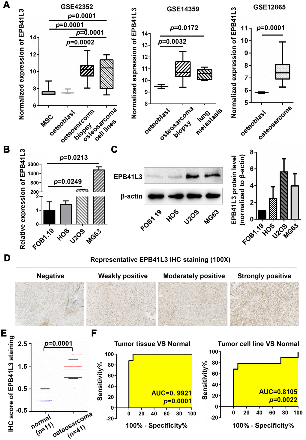
Figure 1.EPB41L3 is up-regulated in osteosarcoma tissues and cell lines. (A) EPB41L3 mRNA expression was significantly higher in osteosarcoma tissues and cell lines than that in normal MSC and osteoblast cells based on data from the Gene Expression Omnibus (GEO accession: GSE42352, GSE14359, and GSE12865). (B) Compared with normal osteoblast cells (hFOB1.19), EPB41L3 mRNA expression was significantly higher in U2OS and MG63 and not obviously up-regulated in HOS as detected by qRT-PCR. GAPDH served as the loading control and was used to normalize the expression data. Data were presented as the mean ± standard error of the mean of two independent experiments. (C) Western blot results showed that EPB41L3 protein expression in U2OS and MG63 but not HOS was significantly higher than that in hFOB1.19 cell. β-actin was used as a loading control and for normalization. Data were presented as the mean ± standard deviation of two independent experiments. (D) IHC staining results of normal bone tissues and osteosarcoma tissues (magnification, x100). (E) IHC total score of EPB41L3 staining was analyzed between normal bone tissues (blue scatter plot, n=11) and osteosarcoma tissues (red scatter plot, n=41). Black solid lines represented the mean ± SD. (F) ROC curves and AUC values for osteosarcoma based on GSE42352. EPB41L3, erythrocyte membrane protein band 4.1-like 3; MSC, mesenchymal stem cell; qRT-PCR, quantitative reverse transcription polymerase chain reaction; GAPDH, glyceraldehyde-3-phosphate dehydrogenase; IHC, immunohistochemistry; ROC, receiver operating characteristic; AUC, area under curve.