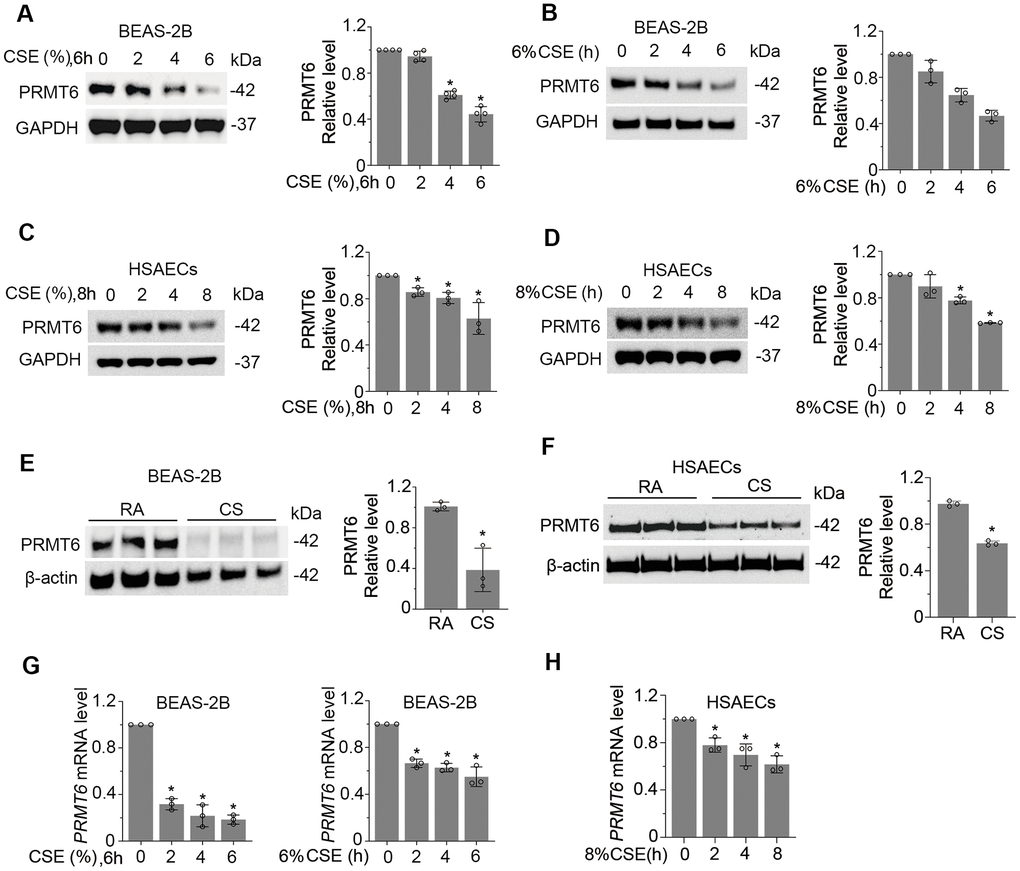
Figure 2.Cigarette smoke reduces the mRNA and protein expression of PRMT6 in airway epithelial cells. (A, B) BEAS-2B cells were treated with CSE in a range of concentrations (A) and time points (B) as indicated. Cell lysates were subjected to immunoblotting for PRMT6 and GAPDH. The densitometric results were plotted in the right panels. (C, D) Human primary small airway epithelial cells (HSAECs) were treated with CSE in different concentrations (C) for 0, 2, 4, 8h (D). Immunoblotting was performed to examine PRMT6 expression. Right panels showed the densitometric results of the blots. (E) BEAS-2B cells were exposed to cigarette smoke. Cell lysates were analyzed with PRMT6 and GAPDH via immunoblotting. RA: room air; CS: cigarette smoke. The densitometry results of the blots were plotted in the right panel. (F) HSAECs cells were exposed with room air (RA) or cigarette smoke (CS). Cell lysate were obtained and analyzed with PRMT6 and GAPDH immunoblotting. Right panels were the plotted densitometric results. (G) Total RNA was extracted from control and CSE-treated BEAS-2B cells (2%, 4%,6% for 6h, and 6% for 0, 2h, 4h, 6h). PRMT6 and GAPDH mRNA levels were determined with qRT-PCR. (H) HSAECs were treated with 8% CSE for 2h, 4h and 8h. Total RNA was extracted and applied to qRT-PCR to detect PRMT6 and GAPDH mRNA level. Relative PRMT6 mRNA level was plotted. Values represent mean ± SD and “*” denotes p < .05. Results were representative of at least n=3 experiments.