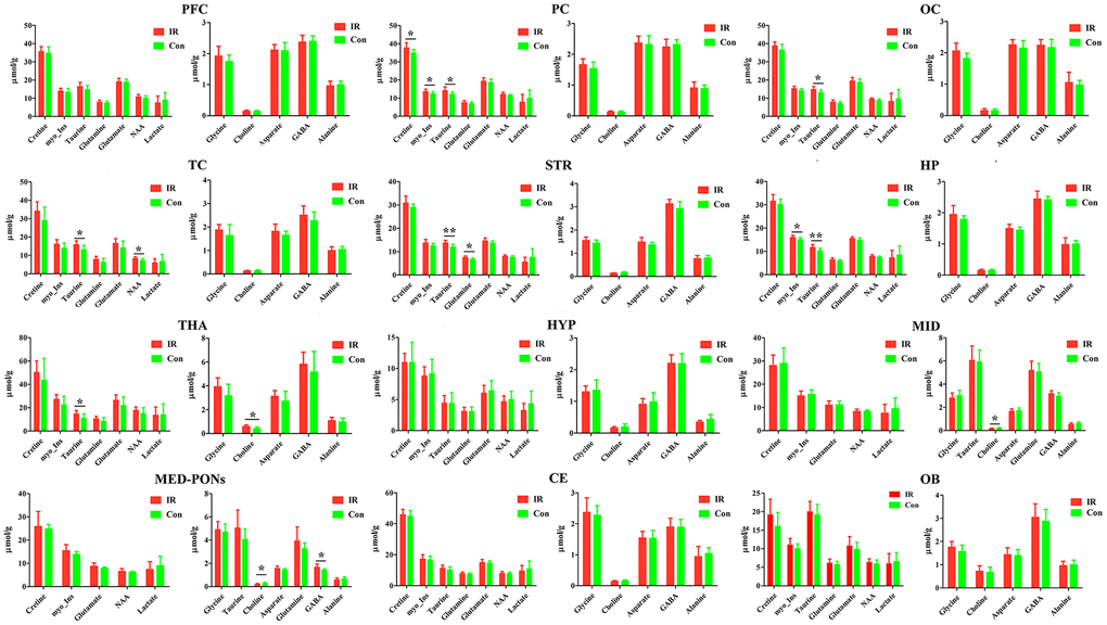
Figure 5.The concentration of identified metabolites in different brain regions in control (con) and ischemia reperfusion (IR) groups using the PMRS method. Data were presented as means ± SEM. Mann-Whitney test. *p< 0.05, **p<0.01. Note: FC, Prefrontal Cortex; PC, Parietal cortex; OC, Occipital cortex; TC, Temporal Cortex; STR, Striatum; HP, Hippocampus; HTA, Thalamus; HYP, Hypothalamus; MID, Midbrain; MED-PONs, Medulla-Pons; CE, Cerebellum; OB, Olfactory Bulbs; GABA, gamma amino acid butyric acid; NAA, N-acetyl aspartate.