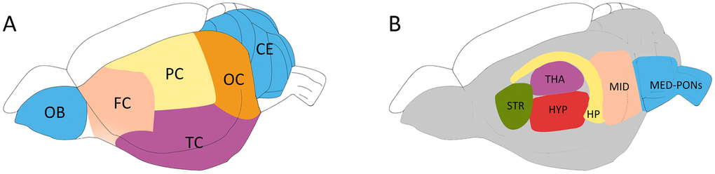
Figure 8.Schematic diagram showing the rat brain regions examined using proton NMR. Blue codes show the olfactory bulb (OB), cerebellum (CE) and cerebral cortical regions (A) and subcortical areas (B) studied. The OB and the cerebellum (CE) were first sampled, followed by the hippocampus (HP), the thalamus (THA) and the striatum (STR). The hypothalamus (HYP), the Midbrain (MID), and Medulla-Pons (MED-PONs) were discarded. The cerebral cortex tissues were coronally cut into four identical parts along the axial axis to represent the frontal cortex (FC), parietal cortex (PC), Temporal cortex (TC), and occipital cortex (OC).