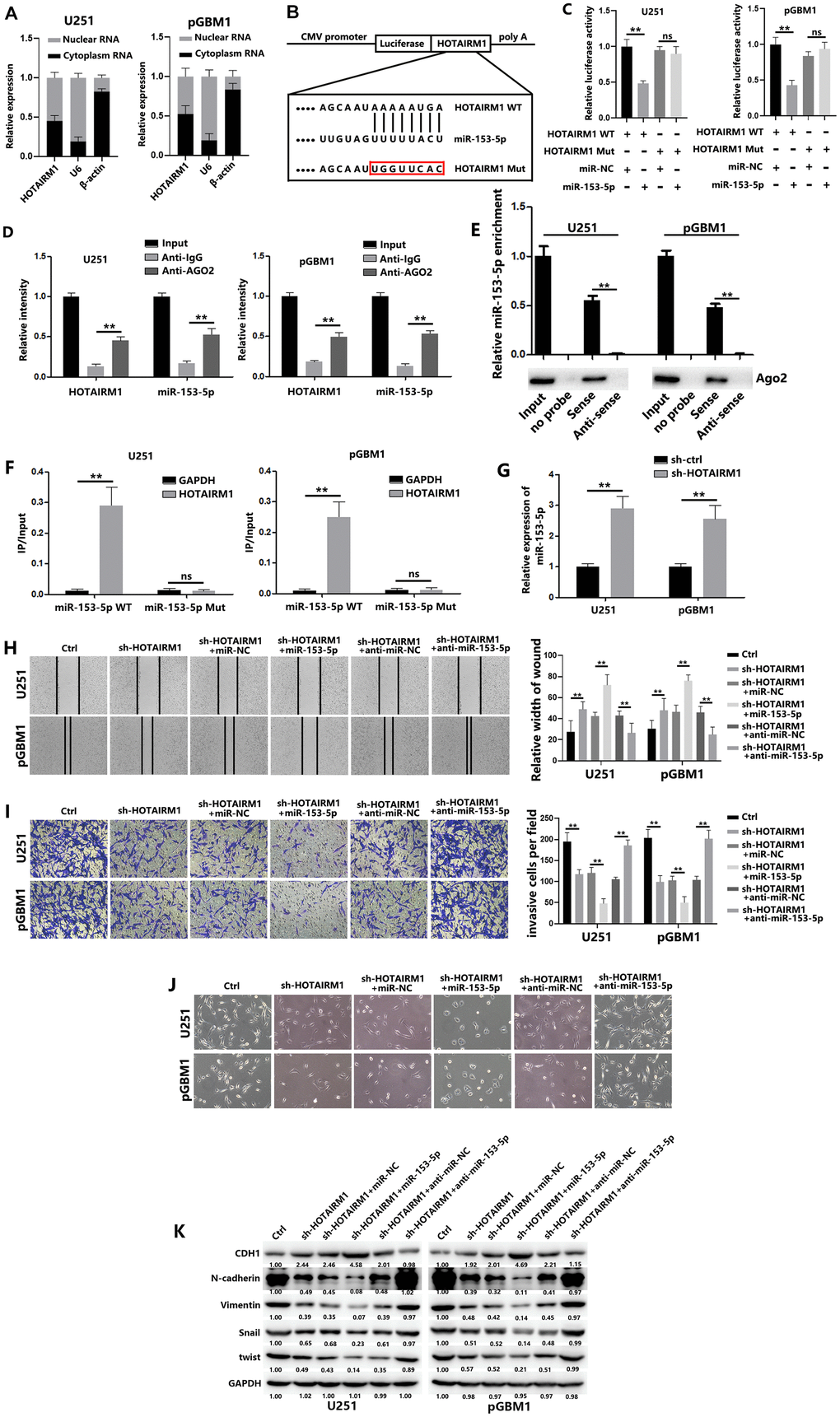
Figure 3.HOTAIRM1 serves as a molecular sponge for miR-153-5p. (A) HOTAIRM1 expression in the nucleus and cytoplasm of GBM cells was measured using qRT-PCR. U6 (nuclear retained) and β-actin (exported to cytoplasm) were used as controls. (B) Schematic diagram shows the putative miR-153-5p binding sites with HOTAIRM1. The sequences of wild-type HOTAIRM1 and mutant HOTAIRM1 are listed as well. (C) Luciferase reporter gene assays were performed to measure the luciferase activity in GBM cells. (D) RNA immunoprecipitation (RIP) assays were performed to determine HOTAIRM1 and miR-153-5p RNA enrichment in immunoprecipitated (IP) complex. Anti-immunoglobulin G (IgG) was used as the control. (E) Relative expression of miR-153-5p and the level of Ago2 in the products of HOTAIRM1 based pull-down assays. (F) The biotinylated miR-153-5p WT or miR-153-5p Mut was transfected into GBM cells. qRT-PCR was performed to quantify the RNA levels of HOTAIRM1 and GAPDH. Relative ratios of the input of IP were analyzed. (G) Relative expression of miR-153-5p in GBM cells was analyzed after transfection with sh-ctrl or sh-HOTAIRM1. (H) Wound healing assays were used to analyze migration of GBM cells. (I) Matrigel invasion assays were used to analyze invasion of GBM cells. (J) Morphological changes of GBM cells were imaged to analyze EMT process of GBM cells. (K) EMT-associated proteins in GBM cells were determined using western blotting. *P < 0.05, **P < 0.01.