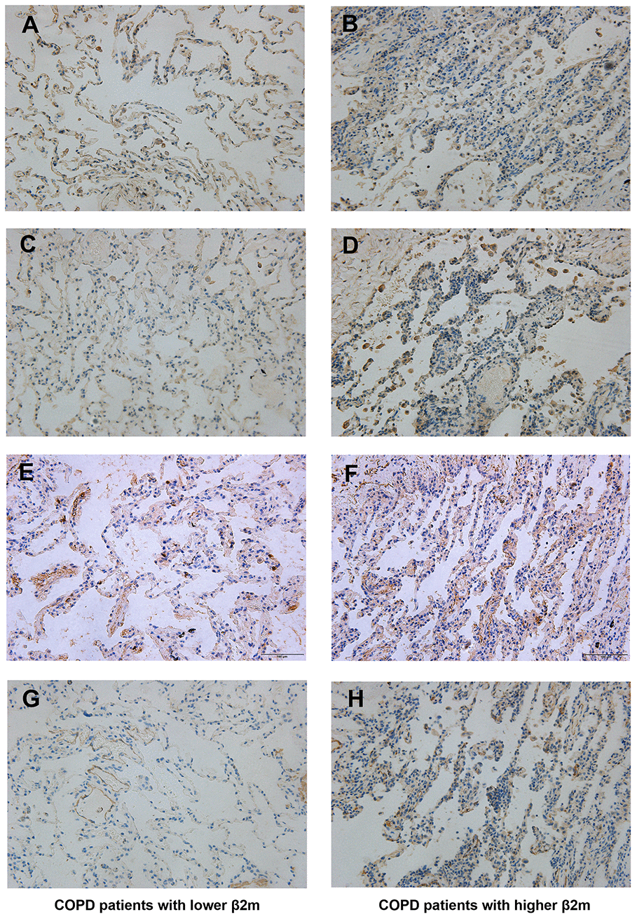
Figure 2.Immunohistochemical staining of lung tissue from COPD patients. Lung tissue of COPD patients with lower serum β2M including (A, C, E, G); lung tissue of COPD patients with higher serum β2M including (B, D, F, H). Indicators and positive cell rate: (A, B) Representative image of β2M immunohistochemical staining of lung tissue from COPD patients with lower serum β2M (17.17 ± 1.64%) and with higher serum β2M (28.95 ± 1.26%) respectively. (C, D) Representative image of TGF-β1 immunohistochemical staining of lung tissue from COPD patients with lower serum β2M (16.48 ± 0.63%) and with higher serum β2M (32.46 ± 0.69%) respectively. (E, F) Representative image of Smad4 immunohistochemical staining of lung tissue from COPD patients with lower serum β2M (34.95 ± 0.71%) and with higher serum β2M (43.38 ± 0.90%) respectively. (G, H) Representative image of a-SMA immunohistochemical staining of lung tissue from COPD patients with lower serum β2M (3.854 ± 0.43%) and with higher serum β2M (26.66 ± 0.89%) respectively. P<0.05 in COPD patients with higher serum β2M versus those with lower serum β2M.