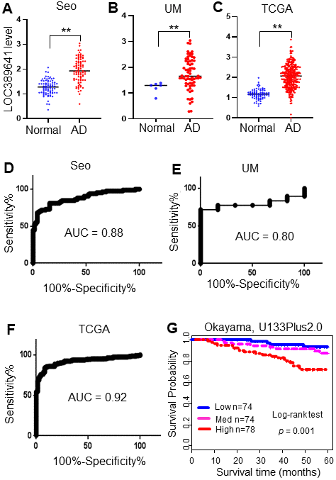
Figure 1.LOC389641 is highly expressed in lung adenocarcinomas and is associated with poorer patient survival. (A–C), Dot plots of LOC389641 expression levels in lung adenocarcinoma (AD) and normal lung (N) tissues in the Seo (85 AD vs 77 N), TCGA (312 AD vs 79 N) and UM (67 AD vs 6 N) RNAseq datasets (y-axis is log2 of FPKM value, ** AD vs. Normal, t test, p < 0.01). (D–F) ROC curves with AUC values of LOC389641 in Seo (85 AD vs 77 N), TCGA (312 AD vs 79 N) and UM (67 AD vs 6 N) RNAseq datasets. (G) Kaplan-Meier survival curve with log-rank test of LOC389641 in Okayama dataset (226 ADs, U133plus2.0 array). Higher LOC389641 expression (1/3 cases for each group based on LOC389641 value) was significantly correlated with poor patient survival.