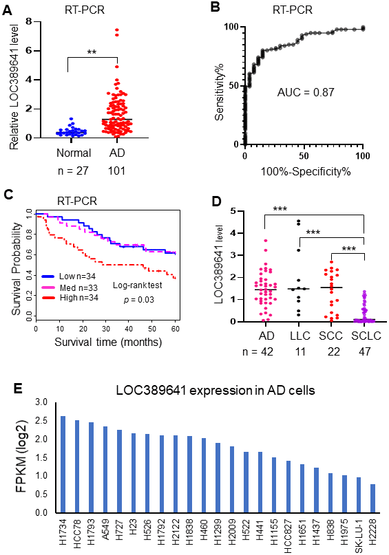
Figure 2.Validation of LOC389641 expression in an independent cohort of ADs and lung cancer cell lines. (A) Dot plot indicating LOC389641 expression was higher in ADs in an independent validation cohort measured by RT-PCR. y-axis is fold-change to mean of all tissues, ** AD vs. normal, t test, p < 0.01. (B) ROC curve indicated an excellent AUC (0.87) for classifying 101 AD from 27 normal lung tissues based on LOC389641 expression in this independent validation cohort. (C) Kaplan-Meier survival curve indicated higher LOC389641 expression was unfavorable for patient survival in this independent validation cohort. Log-rank test, p = 0.03. (D) LOC389641 expression in different types of lung cancer cell lines. LOC389641 expression was significantly lower in small cell lung cancer (SCLC) cell lines (CCLE, RNAseq data, *** NSCLC vs. SCLC, p < 0.001 by t test). (E) LOC389641 expression in individual lung adenocarcinoma cell lines (CCLE, RNAseq data) and these cell lines were ranked in order of LOC389641 expression.