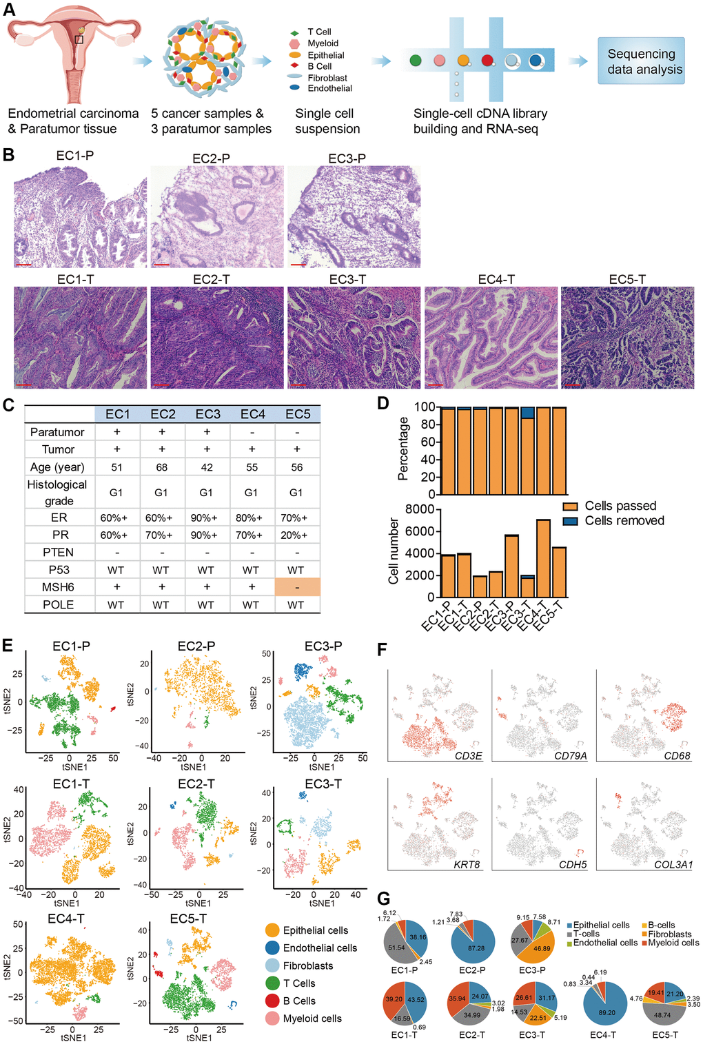
Figure 1.Diversity of cell types in each sample from ECC patients delineated by single-cell transcriptomic analysis. (A) Experimental workflow of scRNA-seq procedure for ECC tumors and paratumor tissues. (B) Hematoxylin and eosin (HE) staining on paratumor and tumor slides of the 5 ECC patients. Scale bars, 100 mm. (C) Samples obtained from 5 EC patients and clinicopathological characteristics of the 5 patients, more details are provided in Supplementary Figure 1B. (D) The remaining cell fraction (left bar plot) and cell number (right bar plot) after quality control and filtering step for each biopsy. (E) The t-distributed stochastic neighbor embedding (t-SNE) plot demonstrates the major cell types in each sample. (F) Expression of representative marker genes of the major cell types defined in EC samples. (G) The percentage of cell types assigned to each sample in (E). Pie charts of cell-type fractions for tumor-infiltrating immune cells of each patient, colored by cell type.