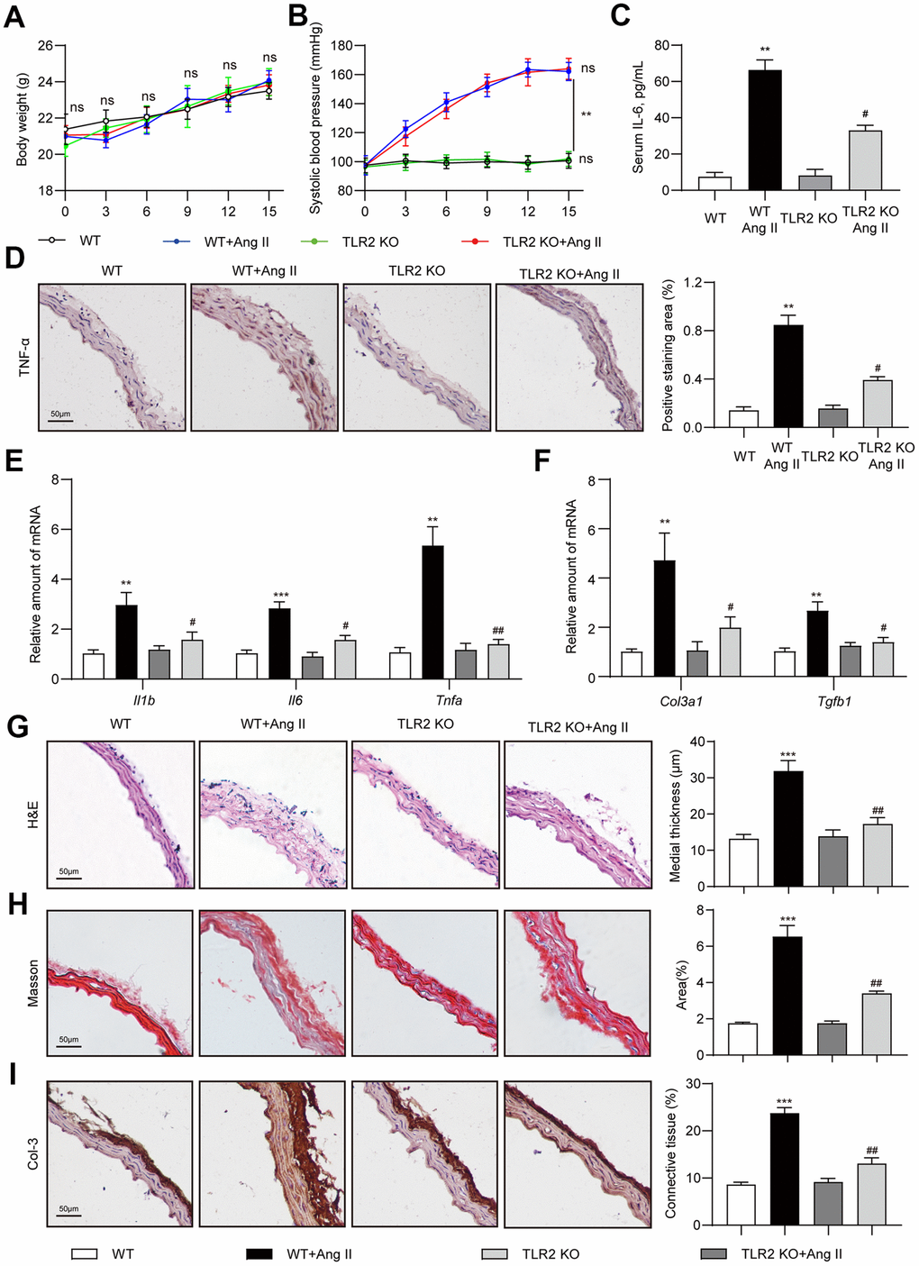
Figure 1.TLR2 deficiency prevents Ang II-induced vascular remodeling in mice. Wildtype (WT) and TLR2 knockout (TLR2KO) mice were challenged with Ang II for 2 weeks. Data showing (A) body weights of mice, (B) systolic blood pressure measurements, and (C) levels of serum interleukin-6 (IL-6). (D) Representative images of TNF-α staining of mouse aortic tissues [scale bar = 50 μm]. Quantification of TNF-α staining area is shown on right. (E, F) mRNA levels of pro-inflammatory and fibrosis-associated genes in aortic tissues [data normalized to β-actin]. (G, H) H&E and Masson's Trichrome staining of mouse aortic sections illustrating the degree of medial thickening and fibrosis [scale bar = 50 μm]. Quantification of medial thickening and connective tissue deposition are shown on right. (I) Representative images of Col-3 staining (brown) of aortic sections. Tissues were counterstained with hematoxylin (blue) [scale bar = 50 μm]. Quantification of Col-3 staining is shown on right. [n = 6-8; Data shown as Mean ± SEM; **p<0.01, and ***p<0.001 compared to WT; #p<0.05 and ##p<0.01 compared to WT-Ang II].