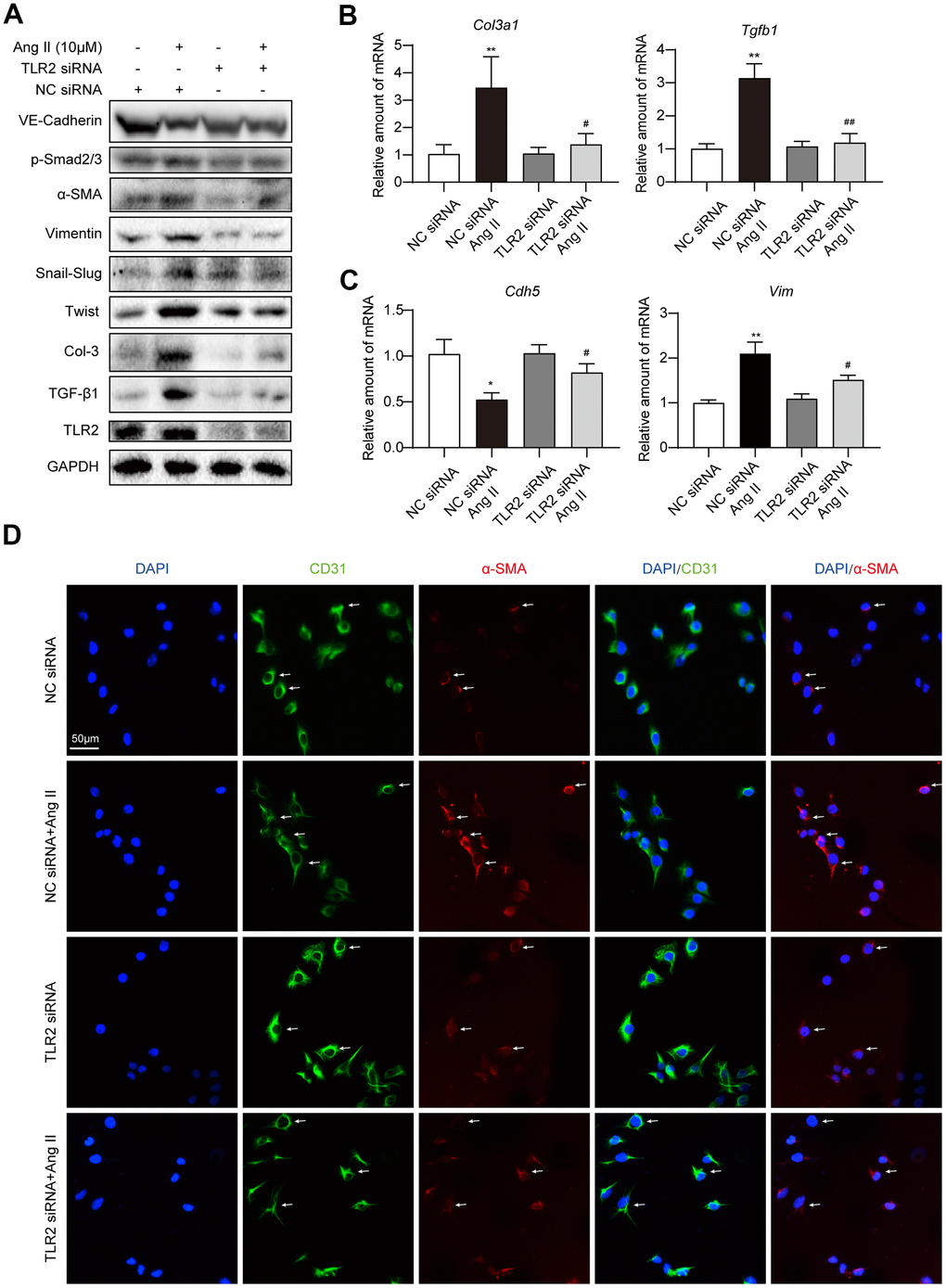
Figure 3.Silencing TLR2 prevents EndMT phenotype in cultured endothelial cells. (A) HUVECs were transfected with siRNA against TLR2. Control cells were transfected with negative control (NC) siRNA. Cells were then challenged with 10 μM Ang II for 36 h. Total proteins were probed for TLR2 and EndMT-associated proteins. GAPDH was used as loading control. (B, C) HUVECs were exposed to 10 μM Ang II for 24 h, after siRNA transfections. mRNA levels of EndMT-associated genes were determined. Data was normalized to β-actin. (D) Representative immunofluorescence staining of HUVECs for CD31 (green) and α-SMA (red). Cells were transfected with indicated siRNA and then exposed to 10 μM Ang II for 36 h. Slides were counterstained with DAPI (blue). Arrows indicate HUVECs undergoing EndMT, as evident through loss of CD31 and induction of α-SMA [Scale bar = 50 μm]. [n = 3; Data shown as Mean ± SEM; *p<0.05 and **p<0.01, compared to negative control transfection; #p<0.05 and ##p<0.01 compared to Ang II challenged negative control transfection].