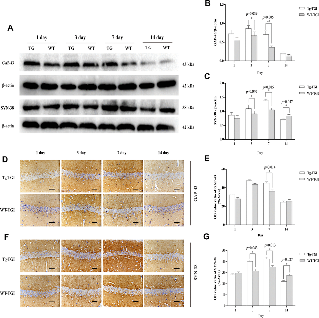
Figure 3.Protective efficacy of neuritin overexpression on cerebral ischemia−reperfusion injury revealed by upregulation of synaptic markers. (A) Western blots of GAP-43 and synaptophysin (SYN-38) expression at the indicated times after transient global ischemia (TGI). β-actin was used as the gel loading control. (B, C) Quantitative analysis of the Western blot results for GAP-43 and SYN-38 shown in (A), respectively. Protein bands were quantified by optical density (OD) measurements. (D, F) Protein expression levels of GAP-43, SYN-38, respectively, in hippocampal CA1 by immunohistochemistry. (E) Quantitative analysis of the immunohistochemistry results for GAP-43 shown in (D). (G) Quantitative analysis of the immunohistochemistry results for SYN-38 shown in (F). Tg-TGI: neuritin-overexpressing transgenic mice subjected to TGI; WT-TGI: wild-type mice subjected to TGI. Six randomly chosen brain sections from three mice were used for statistical analysis. Data are expressed as mean ± S.E.M. n = 6 mice per group, scale bar=50 μm, *p < 0.05, **p < 0.01 versus WT group.