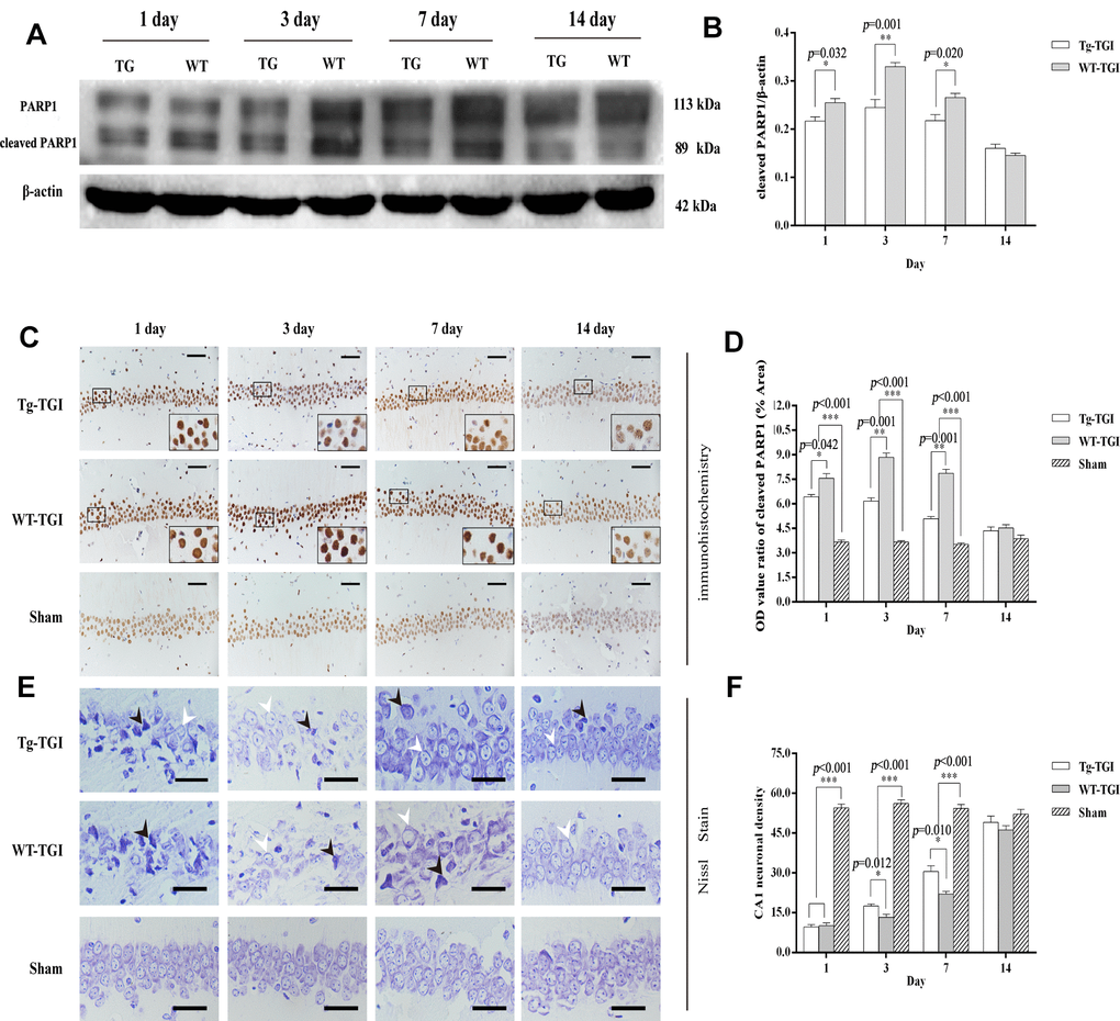
Figure 4.Suppression of TGI-induced apoptosis by neuritin overexpression. (A) Western blots of PARP 1 expression level as an index of apoptosis at the indicated times after TGI. β-actin was used as the gel loading control. Tg-TGI, neuritin transgenic mice subjected to TGI; WT-TGI, WT mice subjected to TGI. (B) Quantitative analysis of cleaved PARP 1 expression data (OD value ratio) shown in (A). (C) PARP 1 protein expression levels in hippocampal CA1 of sham and TGI groups at different time points revealed by immunohistochemistry (scale bar=50 μm). Small black rectangular frame/large black rectangular frame=1/4. (D) Quantitative analysis of expression (OD value ratio) shown in (C). (E) Representative photographs of Nissl-stained hippocampal CA1 at the indicated time points after TGI (scale bar=25 μm). Black arrows indicated the damaged neurons, white arrows indicated the normal neurons. (F) Quantitative analysis of Nissl staining from (E). Cell counts (number of neurons per 200 μm length) from six sections of left and right hippocampus were averaged for each animal. Data expressed as mean ± S.E.M. n = 6 mice per experimental group, *p < 0.05, **p < 0.01 and ***p < 0.001 versus the sham and WT groups, respectively.