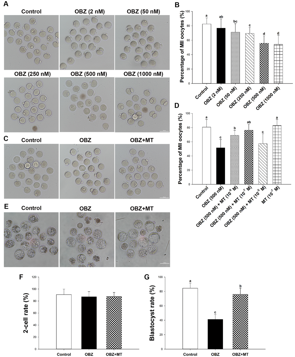
Figure 1.Effects of melatonin on mouse oocytes and embryos following OBZ exposure in vitro. (A) Representative images of oocytes that extruded the first polar body (PB1) in the control and OBZ-exposed groups. Scale bar, 100 μm. (B) Polar body extrusion (PBE) rate in different treatment groups. (C) The mouse oocyte morphologies in the control, OBZ, and OBZ+MT groups. Scale bar, 100 μm. (D) The effects of gradient concentrations of melatonin on the rate of PBE in OBZ-exposed oocytes. (E-G) Mouse embryo morphologies (E) and embryo development rate (F, G) from the 2-cell to blastocyst stages in the control, OBZ, and OBZ+MT groups. Values indicated by different letters are significantly different (P < 0.05). Control, untreated control group; OBZ, oxybenzone-exposed group; OBZ+MT, “oxybenzone + melatonin” treatment group.