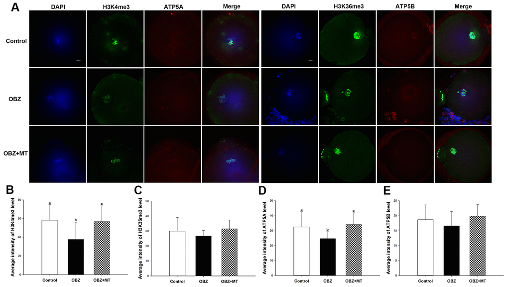
Figure 2.Effects of melatonin on H3K4me3, H3K36me3, ATP5A, and ATP5B levels in OBZ-exposed oocytes. (A) Immunofluorescence staining for H3K4me3, H3K36me3, ATP5A, and ATP5B in the control, OBZ-exposed, and melatonin+OBZ-treated oocytes. Green, H3K4me3 and H3K36me3; red, ATP5A and ATP5B; blue, DNA. Scale bar, 10 μm. (B–E) Average fluorescent intensities for H3K4me3 (B), H3K36me3 (C), ATP5A (D), and ATP5B (E) in mouse oocytes from the different groups. Values indicated by different letters are significantly different (P < 0.05). The experiments were repeated five times, with n = 15-20 per group. Control, untreated control group; OBZ, oxybenzone-exposed group; OBZ+MT, “oxybenzone + melatonin” treatment group.