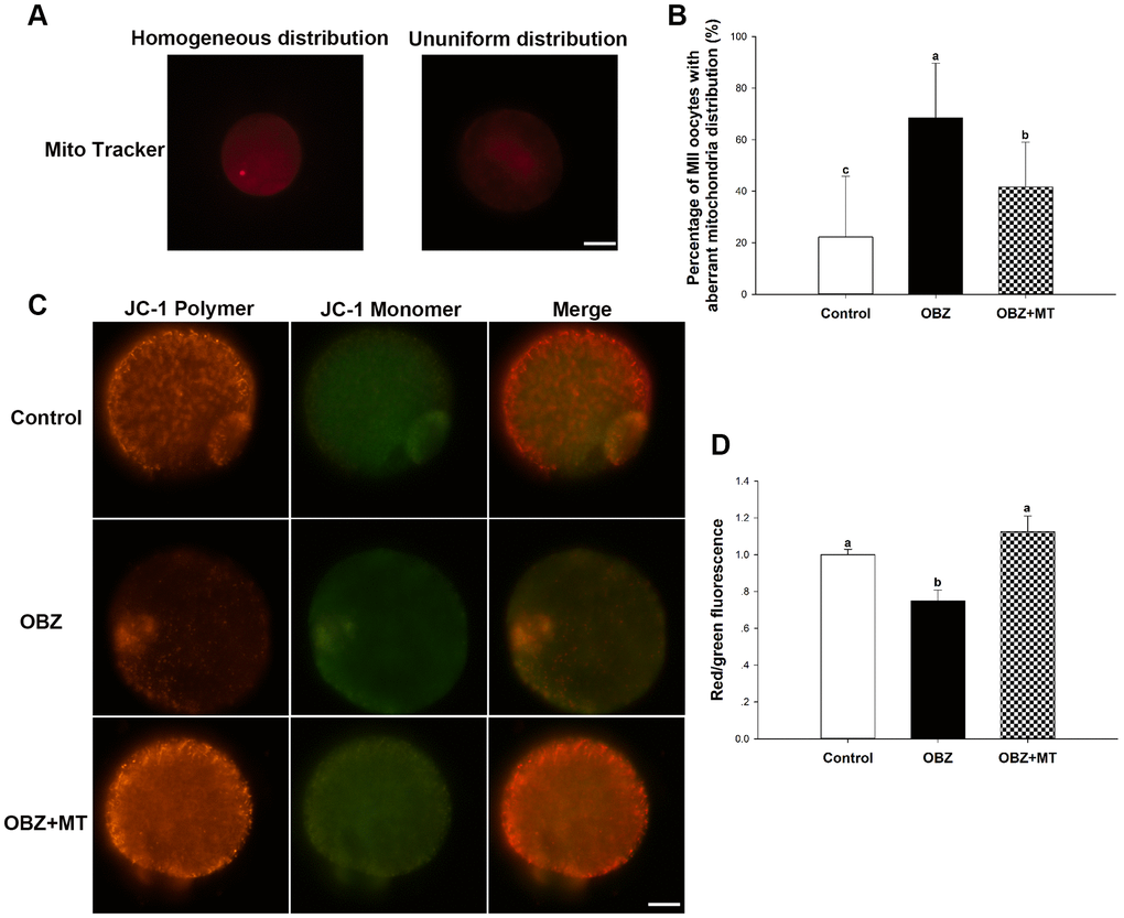
Figure 5.Effects of melatonin on the mitochondrial distribution pattern and mitochondrial membrane potential (ΔΨm) in oxybenzone-exposed mouse oocytes. (A) Representative images of homogeneous and nonuniform distribution patterns of mitochondria in oocytes. Scale bar, 30 μm. (B) Proportion of oocytes with aberrant distribution of mitochondria in each treatment group. (C) Representative images depicting JC-1 in the control, OBZ-exposed, and melatonin+OBZ-treated oocytes. Scale bar, 20 μm. (D) Relative ΔΨm represented as the ratio of red to green intensity. Values indicated by different letters are significantly different (P < 0.05). The experiments were repeated three times, with n = 15-20 per group. Control, untreated control group; OBZ, oxybenzone-exposed group; OBZ+MT, “oxybenzone + melatonin” treatment group.