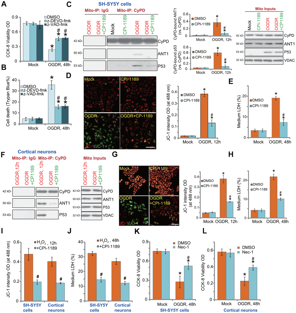
Figure 4.CPI-1189 inhibits OGDR-induced programmed necrosis in neuronal cells. SH-SY5Y cells were pretreated for 1h with z-DEVD-fmk or z-VAD-fmk (each at 50 μM), followed by OGDR stimulation; Cells were cultured for another 48h, cell viability and death were tested by CCK-8 (A) and Trypan blue staining (B) assays, respectively. SH-SY5Y neuronal cells (C–E) or primary murine cortical neurons (F–H) were pretreated for 1h with CPI-1189 (100 nM) and treated with OGDR, cells were cultured for applied time periods, mitochondrial p53-CyPD-ANT1 association (“Mito-IP: CyPD”) and their expression (“Mito Inputs”) were tested (C–F); Mitochondrial depolarization and cell necrosis were tested by JC-1 dye assay (D–G) and medium LDH release (E–H), respectively. SH-SY5Y neuronal cells or primary murine cortical neurons were pretreated for 1h with CPI-1189 (100 nM) and stimulated with hydrogen peroxide (H2O2, 300 μM); Cells were cultured for applied time periods, mitochondrial depolarization (I) and cell necrosis (J) were tested similarly. SH-SY5Y cells or primary cortical neurons were pre-treated for 1 hour with 25 μM of necrostatin-1 (“Nec-1”), followed by OGDR stimulation and cells were then cultured for 48h; Cell viability was tested by CCK-8 assays (K, L). Quantified values were mean ± standard deviation (SD, n=5). * P < 0.05 vs. “Mock” cells. #P < 0.05 vs. cells with OGDR stimulation/H2O2 treatment but “DMSO (0.1%)” pretreatment. Experiments were repeated three times, with similar results obtained. Scale bar= 100 μm (D–G).