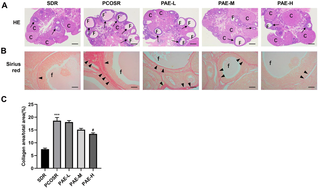
Figure 2.Influence of PAE on ovarian structure, follicle growth and ovarian collagen deposition in PCOS rats. Rats were executed at diestrus. (A) Representative pictures of ovarian tissues in PCOS rats (Hematoxylin and eosin staining, Scale bars = 500 μm, n=10 for each group). (B) Sirius red staining under a light microscope (Scale bars = 100 μm, n=10 for each group). (C) Proportion of the area containing collagen to the ovarian area (mean ± SEM, n=10 for each group). *P < 0.05, **P < 0.01 and ***P < 0.001 vs. SDR group. #P < 0.05, ##P< 0.01 and ###P< 0.01 vs. PCOSR group. PCOS, polycystic ovarian syndrome; SDR, normal control group; PCOSR, PCOS model group; PAE-L, PAE low-dose group (20 mg/kg/d); PAE-M, PAE middle-dose group (40 mg/kg/d); PAE-H, PAE high-dose group (80 mg/kg/d). “▲” is directed in the direction of collagen fibers. “→” is directed in the direction of granular cell layers. C, corpora luteum; F, cyst-like follicles; f, cyst-like follicles.