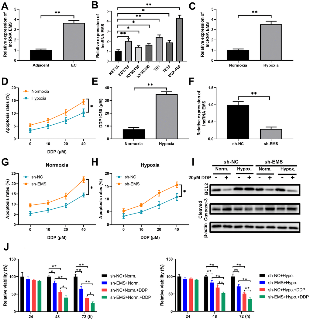
Figure 1.LncRNA EMS is more expressed in esophageal carcinomas and contributes to cancer cell drug resistance against DDP. (A) The expression levels of EMS in esophageal cancer tissues and the adjacent normal tissues were evaluated by RT-qPCR. n=20 for each group. (B) The expression levels of EMS in normal esophageal epithelial HET1A cell and multiple esophageal cancer cell lines (EC9706, ECA-109, KYSE150, KYSE450, TE1 and TE10) were determined by RT-qPCR. n=5 for each group. (C) The expression levels of EMS in ECA-109 cells under hypoxic condition and normoxic condition were determined by RT-qPCR. n=5 for each group. (D) The apoptosis rates (% of Annexin V-positive cells) of ECA-109 cells cultured under normoxic and hypoxic conditions in the presence of DDP at the indicated doses for 24 hours were determined by flow cytometry. (E) ECA-109 cells under hypoxic condition had significantly higher DDP IC50 values than the cells under normoxic condition. (F) qPCR results show the knockdown efficiency of EMS-shRNA in ECA-109 cells. n=5 for each group. (G, H) The apoptosis rates of control ECA-109 cells (sh-NC) and EMS silenced ECA-109 cells (sh-EMS) in response to the indicated concentrations of DDP under normoxic (G) and hypoxic (H) conditions were evaluated by flow cytometry. n=5 for each group. (I, J) Control (sh-NC) and EMS silenced (sh-EMS) ECA-109 cells were treated with 20 μM DDP or the vehicle under normoxic or hypoxic condition, and the BCL-2 and cleaved caspase-3 protein levels at 48 hours after treatments were determined by western blot (I). Cell proliferation under the indicated conditions for 4 days were measured by CCK-8 assays (J). n=5 for each group.