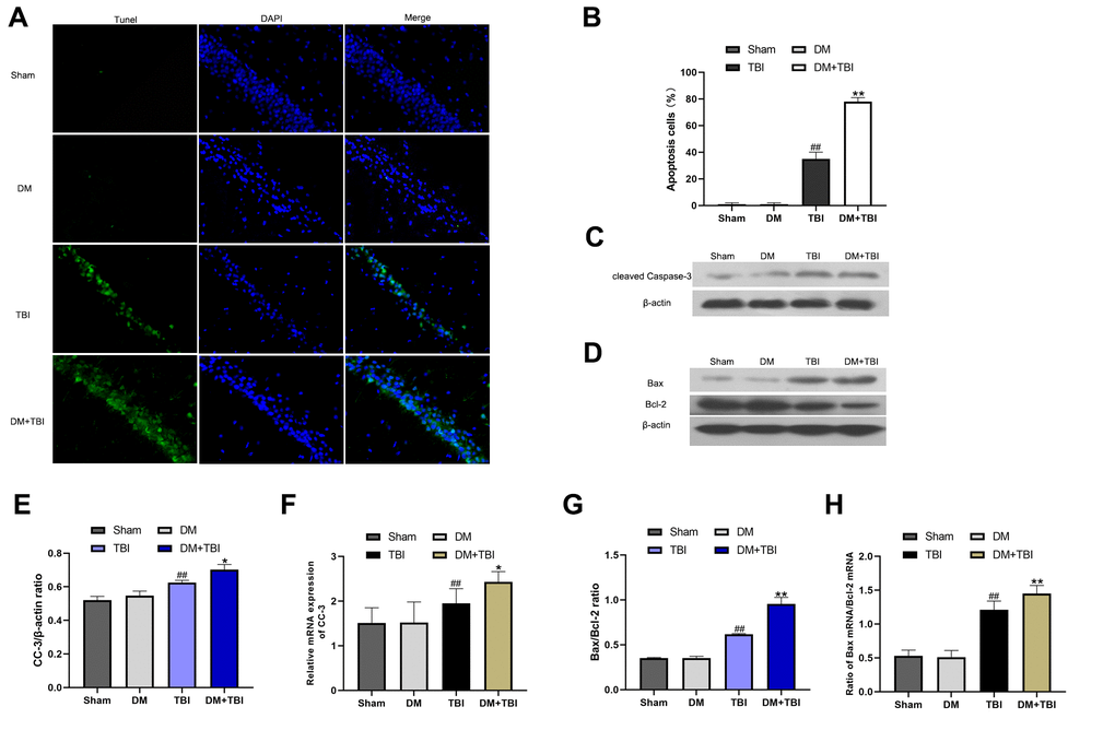
Figure 5.HG exacerbated neuronal apoptosis after TBI. Apoptosis was assessed using DAPI and TUNEL staining 48 h after TBI (scale bar, 50 μm). (A) Representative confocal images of hippocampal tissues stained with TUNEL (green) and DAPI (blue). (B) Bar graph of the proportion of apoptotic cells. Western blot analysis of (C) CC-3 and (D) Bax/Bcl-2 protein levels in the hippocampus 48 h after TBI or sham surgery. Bar graphs illustrate densitometric analyses of the Western blot protein bands for (E) CC-3 and (G) Bax/Bcl-2, each normalized to β-actin. Bar graphs illustrate quantitative analyses of (F) CC-3 and (H) Bax/Bcl-2 mRNA levels, each normalized to β-actin. All data are presented as the mean ± standard error (n = 5 per group). Statistical significance was determined using one-way ANOVA followed by post-hoc Bonferroni correction. #P < 0.05 or ##P < 0.01 vs. the Sham group; *P < 0.05 or **P < 0.01 vs. the TBI group.