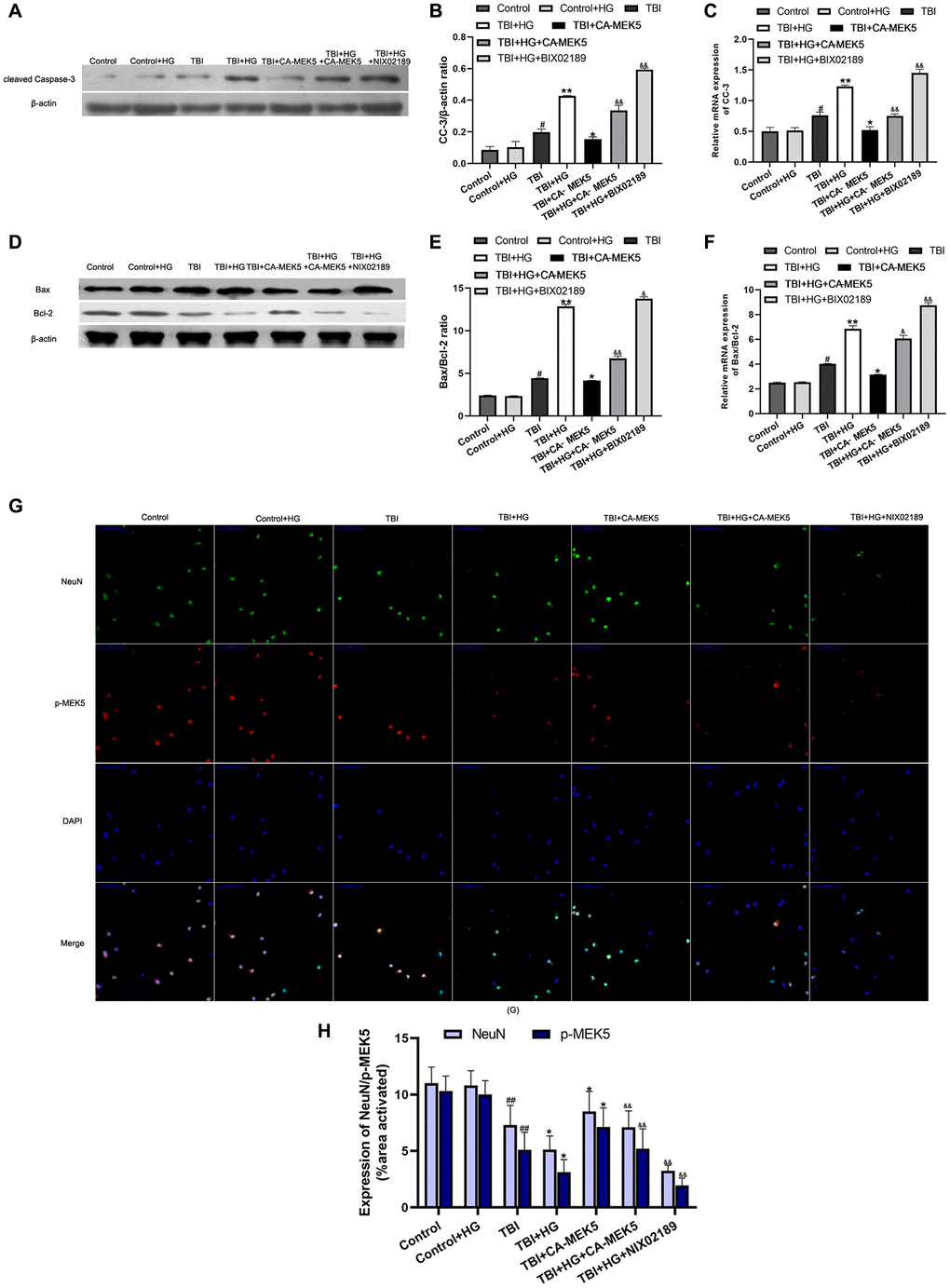
Figure 7.HG increased TBI-induced apoptosis by inhibiting the MEK5/ERK5 pathway. (A) CC-3 and (D) Bax/Bcl-2 protein bands in scratched and transfected primary hippocampal cells. Bar graphs illustrate densitometric analyses of the Western blot protein bands for (B) CC-3 and (E) Bax/Bcl-2, each normalized to β-actin. Bar graphs illustrate quantitative analyses of (C) CC-3 and (F) Bax/Bcl-2 mRNA levels, each normalized to β-actin. (G) Double immunofluorescent staining of NeuN and p-MEK5. Representative confocal images stained for p-MEK5 (red) and NeuN (green) demonstrate that CA-MEK5 treatment not only increased p-MEK5 protein levels, but also markedly increased neuronal survival (scale bar, 100 μm). (H) Staining for NeuN and p-MEK5 was analyzed using MATLAB software. Data are presented as the mean ± standard deviation (n = 5 per group). #P < 0.05 vs. the Control group; *P < 0.05 or **P < 0.01 vs. the TBI group; &P < 0.05 or &&P < 0.01 vs. the TBI+HG group.