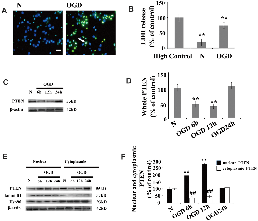
Figure 1.OGD increases PTEN nuclear translocation in cultured neurons (A) TUNEL-positive cells increased 12 h after OGD. The arrow indicates TUNEL-positive cells with green fluorescence. Scale bars = 50 μm. (B) Extracellular LDH levels were elevated at 12 h after OGD. (C) The PTEN whole cell protein levels decreased between 6 h and 12 h after OGD. (D) Quantification of the whole cell protein levels of PTEN. (E) Western blot analysis of the cytoplasmic and nuclear fractions showed that nuclear PTEN significantly increased 6 h after OGD, reaching a peak at 12 h, and returned to control levels within 24 h. Blots were re-probed using cytoplasmic and nuclear protein markers, Hsp90 and lamin B1, respectively. (F) Quantification of PTEN protein levels in cytoplasmic and nuclear fractions. Data were quantified by densitometry and normalized against healthy neurons. n = 5 for each column. **p < 0.01 vs. normal neurons. ## p < 0.01 vs. normal neurons. OGD: oxygen and glucose deprivation; N, normal neurons; TUNEL, terminal deoxynucleotidyl transferase-mediated dUTP-biotin nick-end labeling.