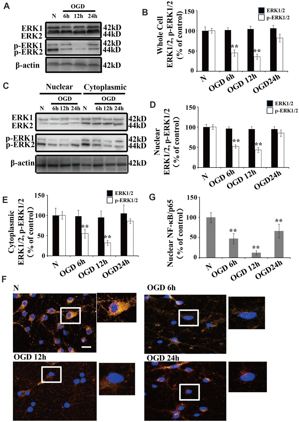
Figure 2.OGD inhibits the phosphorylation of ERK1/2 and NF-κB activation. (A) Whole cell protein levels of total ERK after OGD. The expression of p-ERK1/2 in whole cell extracts was significantly decreased 6 h after OGD, peaked at 12 h, and recovered at 24 h. (B) Quantification of ERK1/2 and p-ERK1/2 expression in whole cell extracts, normalized against normal neurons. (C) The expression of p-ERK1/2 in cytoplasmic and nuclear fractions. Western blot shows that nuclear p-ERK1/2 significantly decreased 6 h after OGD, peaked at 12 h, and recovered at 24 h. The changes in p-ERK1/2 expression in the cytoplasm was similar to that of the nucleus. (D) Quantification of nuclear ERK1/2 and p-ERK1/2 expression, normalized against normal neurons. (E) Quantification of cytoplasmic ERK1/2 and p-ERK1/2 expression, normalized against normal neurons. (F) Immunofluorescence staining of nuclear NF-κB. The nuclear translocation of NF-κB decreased after OGD, with minimal presence in the nucleus at 12 h post treatment. The arrow indicates nuclear-NF-κB/p65-positive cells with red fluorescence. (G) Qualification of fluorescence intensity of nuclear NF-κB staining, normalized against normal neurons. n = 5 in each column and **p < 0.01, vs. normal neurons. OGD: oxygen and glucose deprivation.