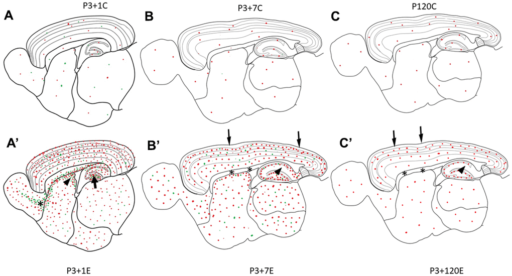
Figure 4.Diagrams (A–C) show γH2AX foci (red dots) in the brain of the control animal without irradiation. In P3+1C or P4 mouse brain, a few apoptotic bodies (green dots) randomly appear in different brain regions (A). One day after irradiation at P3, γH2AX foci (red dots) and apoptotic bodies (green dots) increase obviously in all brain regions (A’). Drastic increase of apoptotic bodies (green dots) appear in the hilus of the dentate gyrus (arrow), in the subventricular zone of the lateral ventricle (arrowhead) and in the rostral migratory stream (asterisk) (A’). Seven days after irradiation at P3, many γH2AX foci (red dots) and apoptotic bodies (green dots) still exist in all brain regions (B’). However, there was no obvious change of γH2AX foci in the pia mater (arrow), white matter (corpus callosum, asterisks), the strata laculosum moleculare, radiatum (arrow), oriens of CA1-3 areas, the stratum moleculare of the dentate gyrus (B’). One hundred-twenty days after irradiation at P3, there are still some γH2AX foci (red dots) still exist in all brain regions (C’) although the number of γH2AX foci is reduced obviously.