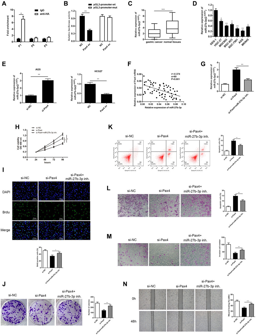
Figure 4.PAX4 targets and negatively regulates miR-27b-3p, and miR-27b-3p reversed PAX4 function in AGS cells. (A, B) The interaction between PAX4 and miR-27b-3p promoter region was validated through ChIP (P = 0.014) and dual luciferase assays (P = 0.0012). (C) The expression of miR-27b-3p was down-regulated in GC tissues (n = 60, P = 0.0004). (D) The expression of miR-27b-3p was down-regulated in six GC cell lines (HGC-27, MGC803, BGC-823, SGC-7901, AGS, MKN45) compared to the human gastric mucosal epithelial cell line GES-1. (E) Up-regulation of miR-27b-3p level was detected in the PAX4 knockdown group in AGS cells (P = 0.0061) and down-regulated miR-27b-3p levels were detected in the PAX4 overexpression group in HGC-27 cells by qRT-PCR (P = 0.0095). (F) There is a negative correlation between PAX4 and miR-27b-3p expression. (G) Lower miR-27b-3p levels were detected via qRT-PCR after addition of miR-27b-3p inhibitor to the si-PAX4 group (P = 0.0077). (H–I) CCK-8 and BrdU assays (P = 0.0035) detected that decreased miR-27b-3p level promoted enhanced GC cell viability and proliferation abilities. (J) Colony formation assay determined the numbers of colony formation (P = 0.0059). (K) GC cell apoptosis was examined via flow cytometry assay (P = 0.0077). (L–M) The increased cell migration (P = 0.0051) and invasion (P = 0.0051) abilities were identified when miR-27b-3p expression was inhibited by transwell assays. (N) Wound healing assay was utilized to discover enhanced GC cell metastasis in the absence of miR-27b-3p compared to the si-PAX4 group (P = 0.0006).