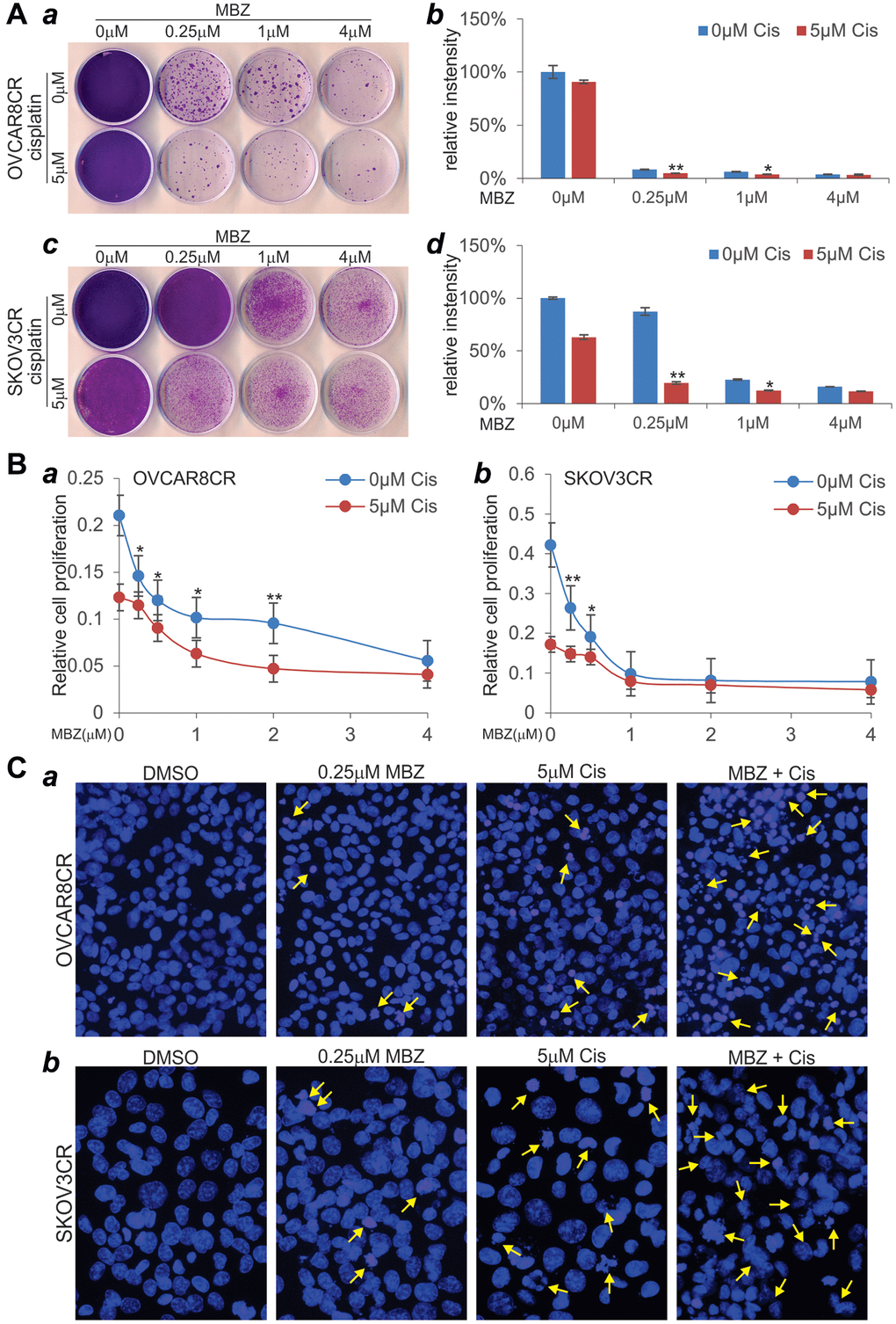
Figure 5.MBZ synergizes with cisplatin to inhibit cell proliferation and induce apoptosis in the human CR ovarian cancer cells. (A) Colony formation and crystal violet cell viability assay. Subconfluent OVCAR8CR (a, b) and SKOV3CR (c, d) cells were treated with MBZ and cisplatin at the indicated concentrations. At 72 h post treatment, the cells were replated for colony formation for 10 days, followed by crystal violet staining (a, c). Each assay condition was done in triplicate. Representative results are shown (a, c). The stained cells were dissolved in acetic acid and quantitatively measured for optical absorbance (b, d). *p < 0.05 **p < 0.01, when compared with that of the respective 0 μM cisplatin groups. (B) WST-1 cell proliferation assay. Subconfluent OVCAR8CR (a) and SKOV3CR (b) cells were seeded into 96-well cell culture plates, and treated with DMSO, cisplatin and/or MBZ at the indicated concentrations. At 72 h post treatment, WST-1 working mix was added to each well and incubated for 2h prior to absorbance reading at 450nm. Each assay condition was done in triplicate. *p < 0.05 **p < 0.01, when compared with that of the respective 0 μM cisplatin groups. (C) Cell apoptosis assay. Subconfluent OVCAR8CR (a) and SKOV3CR (b) cells were seeded into 6-well cell culture plates, and treated with DMSO, 5 μM cisplatin (Cis) and/or 0.25 μM MBZ. At 72 h, the cells were collected, fixed, stained with Hoechst33258, and examined under a fluorescence microscope. Representative images are shown. Representative apoptotic cells are indicated by arrows.