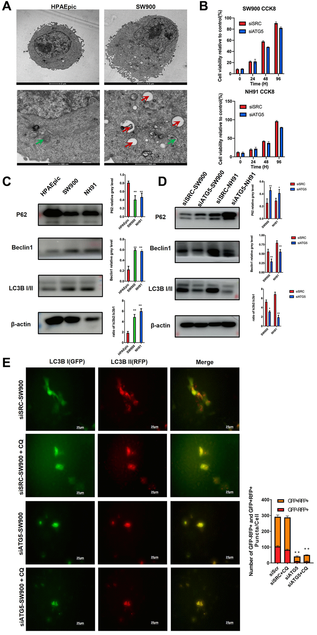
Figure 2.ATG5 down regulation inhibits autophagy in LUSC cells. (A) Representative TEM images showing the formation of autophagosomes (red arrow) and autolysosomes (green arrow) in HPAEpic and SW900. (B) After the transfected with siATG5/SRC, cell viability at different time points were assessed by using an MTT assay. (C) Expression of autophagy relative protein, including P62, Beclin1 and LC3B I/II, were evaluated in HPAEpic, SW900 and NH91cells. (D) Expression of ATG5, P62, Beclin 1, and LC3B I/II in SW900 and NH91 cells transfected with si-ATG5 were detected using a western blot assay. (E) siATG5 and siSRC SW900 cells were transfected with GFP-mRFP-LC3 virus and incubated with or without chloroquine for 24 h and then starved for 2 hours. Red or yellow puncta indicating complete/incomplete autophagy were calculated and reported as the mean ± SD. All the P values were compared with the control *P < 0.05, **P < 0.01, ***P < 0.0001.