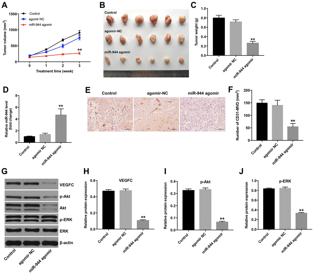
Figure 8.Overexpression of miR-944 inhibits in vivo xenograft glioma cell tumor growth and angiogenesis. (A) The line graph shows tumor volumes on weeks 1-3 in control, agomiR-NC, and agomiR-944 group nude mice. (B) The photographs show xenograft glioma cell tumors harvested at four weeks from control, agomiR-NC, and agomiR-944 group nude mice. (C) The histogram shows the weights of xenograft glioma cell tumors harvested at four weeks from control, agomiR-NC, and agomiR-944 group nude mice. (D) RT-qPCR analysis shows miR-944 levels in the xenograft glioma cell tumor tissues harvested from nude mice belonging to the control, agomiR-NC, and agomiR-944 groups. (E, F) IHC staining results show CD31-stained xenograft glioma cell tumor tissues harvested from nude mice belonging to the control, agomiR-NC, and agomiR-944 groups. Microvessel density (MVD) was analyzed based on CD31+ve staining. (G–J) Western blot analysis shows the levels of VEGFC, p-Akt, Akt, p-ERK, and ERK proteins in the xenograft glioma cell tumor tissues harvested from control, agomiR-NC, and agomiR-944 groups. VEGFC, p-Akt, and p-ERK levels were normalized to β-actin, Akt and ERK levels, respectively. **P < 0.01 vs. the agomiR-NC group; NC, negative control; MVD, microvessel density; IHC, immunohistochemistry.