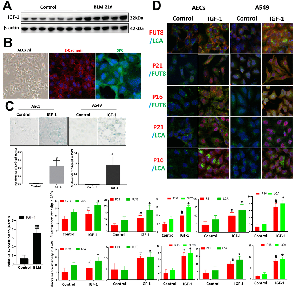
Figure 3.Core fucosylation was increased during the IGF1-induced AEC senescence in vitro. Primary cultures of AECs and A549 cells were incubated with IGF-1 (10 ng/ml) for 72 h. (A) The level of IGF1 was assessed using western blotting in different groups. (B) Representative bright-field images in AECs, and representative images of E-cadherin (red), SPC (green) staining is shown (original magnification, 200×). (C, D) SA-β-gal staining and representative images of dual staining for LCA (green) and FUT8 (red), FUT8 (green) and P21 (red), FUT8 (green) and P16 (red), LCA (green) and P21 (red) and LCA (green) and P16 (red) were performed to detect cellular senescence (original magnification, 200×). Data are shown as the mean ± SEM, n ≥ 3 per group. ##P < 0.01 for the comparison between the control group and the BLM group. *P < 0.01, #P < 0.01 for the comparison between the control group and the IGF1 group. Unpaired, two-tailed Student’s t test.