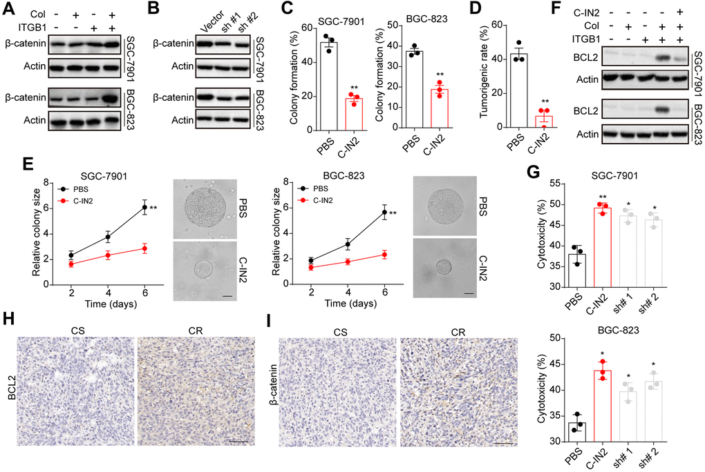
Figure 4.BCL9L mediated gastric progression through downstream β-catenin and BCL2. (A) ITGB1-/+ SGC-7901/BGC-823 cells were sorted and cultured with 3D collagen gel (6 days) or not. The expression of β-catenin and β-actin was examined by western blotting. (B) ITGB1+ SGC-7901/BGC-823 cells were cultured in 3D collagen gels (6 days) and treated with BCL9L shRNA or vector. Then the expression of β-catenin and β-actin was examined by western blotting. (C) ITGB1+ SGC-7901/BGC-823 cells were cultured in 3D collagen gels (6 days) and treated with PBS or C-IN2 (5 μM). Then the 3D colony formation capability was examined. (D) ITGB1+ SGC-7901/BGC-823 cells were cultured in 3D collagen gels (6 days) and treated with PBS or C-IN2 (5 μM). Then the 3D colony formation capability was examined. Then the tumorigenic capability was examined in NOD-SCID mice. (E) ITGB1+ SGC-7901/BGC-823 cells were cultured in 3D collagen gels (6 days) and treated with PBS or C-IN2 (5 μM). Then the colony sizes were examined. The scale bar is 30 μm. (F) ITGB1-/+ SGC-7901/BGC-823 cells were cultured in 3D collagen gel (6 days) and treated with PBS or C-IN2 (5 μM). Then the expression of BCL2 and β-actin was examined by western blotting. (G) ITGB1+ SGC-7901/BGC-823 cells were cultured in 3D collagen gel (6 days) and treated with PBS, C-IN2 (5 μM) or BCL2 shRNA. Then tumor cells were treated with 5-FU (5 μg/ml) and the apoptosis was examined. (H) Immunohistochemical staining of β-catenin in chemo-sensitive (CS) and chemo-resistant (CR) tissues from gastric patients. The scale bar is 100 μm. (I) immunohistochemical staining of BCL2 in chemo-sensitive (CS) and chemo-resistant (CR) tissues from gastric patients. The scale bar is 100 μm. * Indicates P <0.05, ** Indicates P <0.01.