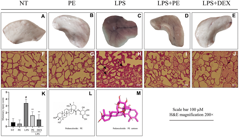
Figure 1.Effect of Pedunculoside (PE) on the pathological damage of mammary gland in mice with mastitis. PE (10 mg/kg) was given orally for 7 days before the establishment of mastitis model. The fourth pair of milk ducts in mice were injected with 50 μL of 0.2 μg/μL LPS for 24 h. The mice were killed by dislocation and fixed on the operating platform. The hair was disinfected and fixed by spraying 70% alcohol. The midline of abdomen was cut to expose the breast tissue. Finally, the mammary was photographed and collected. (A–E) Morphological photos of mouse mammary gland tissue; (F–J) H&E staining of mouse mammary gland paraffin section; (K) Pathological damage score of mice mammary gland tissue; (L) Structural formula of Pedunculoside; (M) 3D mode of Pedunculoside. The lesion was shown by the arrow in the figure. Scale bar 100 μM, H&E magnification 200×. Dexamethasone (DEX) was administered intramuscularly at a concentration of 5 mg/kg. Values are presented as means ± SEM, three independent repeated experiments were performed; #p<0.01 vs. No treatment group (NT) group; **p<0.01 vs. LPS group.