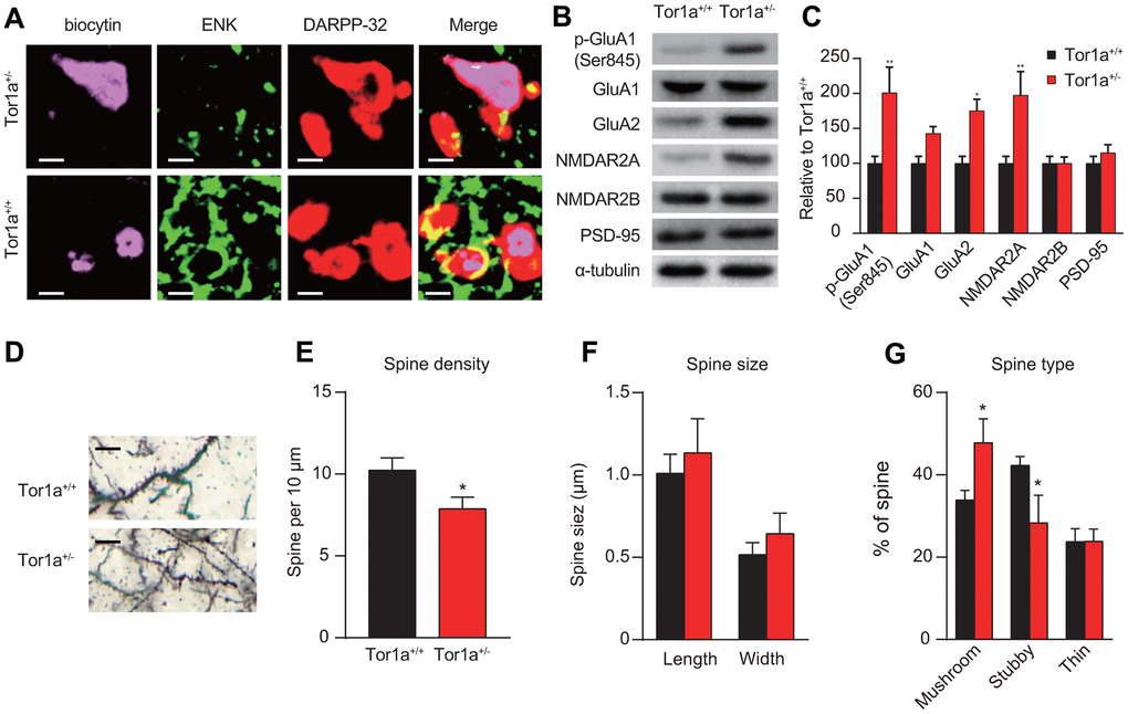
Figure 2.Altered molecular markers and morphology of synapse in Tor1a+/- mice. (A) Representative confocal images from two SPNs recorded in Tor1a+/- and Tor1a+/+ slices (scale bar: 10 μm). Recording electrodes were filled with biocytin (pink) and SPNs were immunolabelled with anti-ENK (green) and anti-DARPP-32 (red). ENK-negative SPNs from Tor1a+/- slices failed to induce LTD. (B) WB analysis for p-GluA1 (Ser845), GluA1, GluA2, NMDAR2A, NMDAR2B, PSD-95 and α-tubulin in Tor1a+/- and age-matched Tor1a+/+ mice. (C) Histogram showed the quantification of protein levels following normalization on α-tubulin in Tor1a+/- and age-matched Tor1a+/+ mice. All values are mean ± SEM expressed as % of Tor1a+/+ mice. (D) Representative images showed spine morphology of Tor1a+/- and age-matched Tor1a+/+ mice (scale bar: 10 μm). (E) Histogram represented dendritic spine density in Tor1a+/- and Tor1a+/+ SPNs. Tor1a+/- SPNs exhibited an overall decrease of dendritic spine density (P<0.05). (F, G) Histograms showed the quantification of dendritic spine size (F, spine length and head width) and dendritic spine type (G, mushroom, stubby, thin) in Tor1a+/- and age-matched Tor1a+/+ mice. A larger number of mushroom-type spines and a concomitant smaller number of stubby-type spines were found in Tor1a+/- SPNs (both P<0.05). In each group, five mice were used (N=5), and three independent experiments were conducted for each mouse (n=3). P<0.05 was considered to be statistically significant.