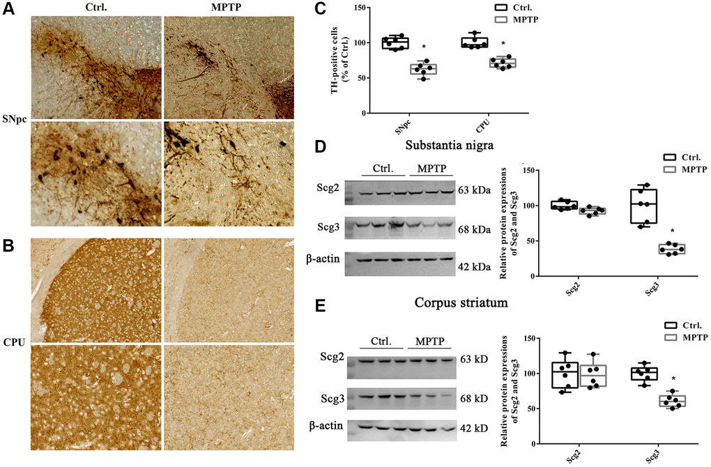
Figure 1.Immunohistochemistry of tyrosine hydroxylase positive neurons and levels of secretogranins in the substantia nigra and corpus striatum. (A–B) Tissues were immunostained for tyrosine hydroxylase (TH) in both cell bodies in substantia nigra pars compacta (SNpc) and fibers and terminals in the corpus striatum (CPU) in MPTP-treated and control groups. (C) (TH)–positive stains in MPTP-treated and control groups were assessed by mean optical density via Image J software. (D–E) Scg2 and Scg3 protein expressions in SN and CPU were analyzed by immunoblotting and quantified by densitometric analysis normalized to GAPDH. Two-tailed unpaired Student t-tests were performed between the control and treated groups. *Statistically significant with P < 0.05; Error bars are SD; N = 6.