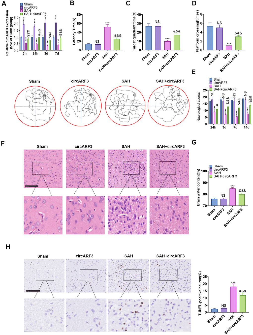
Figure 2.Overexpressing circARF3 attenuated nerve function injury and neuronal apoptosis in SAH rats. The circARF3 overexpression rat model was constructed using lentivirus-carried circARF3 vectors, and the SAH rat model was also constructed. (A) The circARF3 expression in rat brain tissues at different time points (3 hours, 24 hours, 3 days, and 7 days) was verified by RT-PCR. (B–D) On the 16th day of SAH, the water maze experiment was conducted to evaluate the learning and memory function of rats. (E) The modified Garcia score was applied to assess the motor function in rats. (F, G) Changes of cerebral edema were determined by HE staining (F) and wet/dry method (G). (H) TUNEL assay was implemented to detect the apoptosis rate of neurons in brain lesions. NS, ***P>0.05, P<0.001 vs. Sham group. &, &&&P<0.05, P<0.001 vs. SAH group. N=10. Scale bar=100 μm.