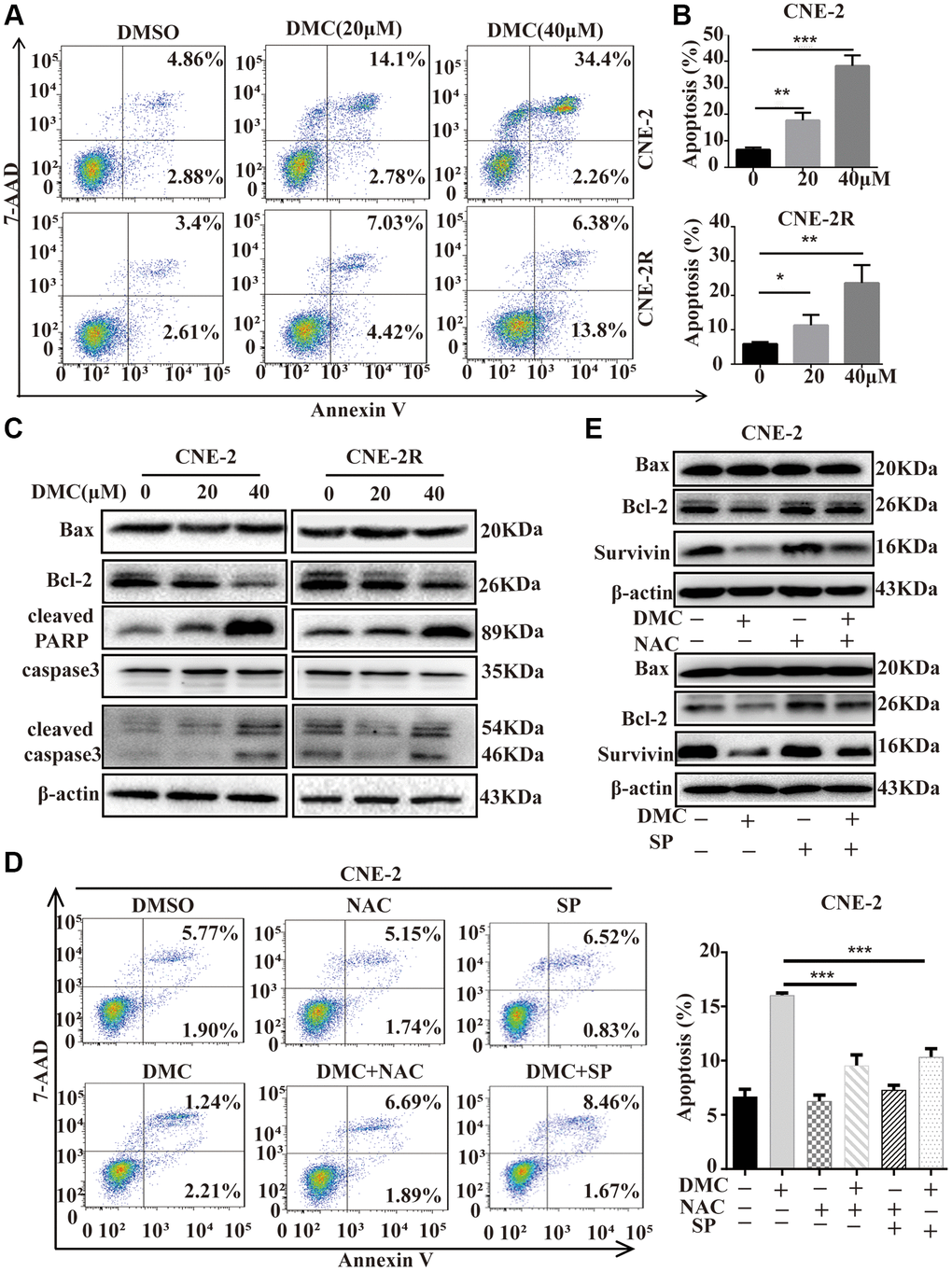
Figure 5.DMC induced apoptosis and autophagic death in NPC cells. (A) CNE-2 and CNE-2R cells were treated with 0.1% DMSO or DMC (20 and 40 μM) for 48 h, and then apoptotic cells were detected with the annexin V-PE/7-AAD kit and analysed by flow cytometry. (B) Statistical analysis histogram of apoptotic cells ratio. (C) Cells were treated with DMC 0.1% DMSO or DMC (20 and 40 μM) for 48 h, and the expression levels of apoptosis-related proteins (cleaved-PARP, cleaved-caspase-3, caspase-3, Bax, and Bcl-2) were tested in cells by Western blotting. β-actin was used as a normalization control. (D) CNE-2 cells were treated with 0.1% DMSO or 20 μM DMC for 24 h in the absence or presence of NAC (5 mM) pretreatment for 1h or SP600125 (30 μM) pretreatment for 1 h, and then apoptotic cells were detected by flow cytometry. Statistical analysis histogram of apoptotic cells ratio. (E) The expression levels of Bax, Bcl-2 and Survivin were tested in CNE-2 cells as Figure 5D by Western blotting. **P < 0.01 and ***P < 0.001, significantly different compared with control group.