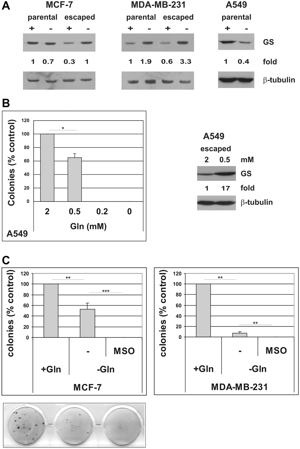
Figure 7.Induction of GS mediates resistance to glutamine deprivation in cells that escape from TIS. (A) Expression of GS protein was analyzed in parental MCF-7, MDA-MB-231 and A549 cells, grown in the presence or in the absence of glutamine for 72 hours, and in escaped clones arisen in the presence or in the absence of glutamine. Filters were stripped and reprobed with anti-β-tubulin antibodies as a loading control. GS levels, normalized to the relative β-tubulin levels, are reported as fold change of Gln-supplemented parental cells. (B) Left panel: doxorubicin-induced senescent A549 cells were grown in media with different glutamine concentrations. Colonies that evaded the senescent growth arrest were stained and counted. Data are mean ± S.D. of two independent experiments. Right panel: expression of GS protein was analyzed in A549 escaped clones arisen in the presence of 2 mM or 0.5 mM glutamine. Filters were stripped and reprobed with anti-β-tubulin antibodies as a loading control. GS levels, normalized to the relative β-tubulin levels, are reported as fold change of Gln-supplemented sample. (C) Inhibition of GS with MSO abolishes escape from TIS in Gln-deprived conditions. Doxorubicin-induced senescent MCF-7 and MDA-MB-231 cells were grown in complete medium (+Gln) or in Gln-deprived medium (−Gln), in the presence or in the absence of 2 mM MSO. Colonies that evaded the senescent growth arrest were stained and counted. Data are mean ± S.D. of three independent experiments.