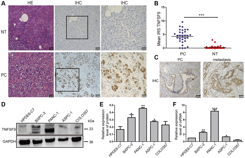
Figure 1.The expression of TNFSF9 in pancreatic cancer tissues is significantly higher than that in non-tumor tissues adjacent to cancer. (A) Representative HE staining and immunohistochemical staining pictures of cancer tissues and non-tumor tissues adjacent to pancreatic cancer (n = 30) and immune response score (B). (C) Immunohistochemical staining pictures of pancreatic cancer tissue with metastasis and pancreatic cancer tissue without metastasis. (D, E) Western blot analysis of the protein expression of TNFSF9 in pancreatic cancer cells COLO357, ASPC-1, PANC-1 and BXPC-3 and normal pancreatic epithelial cells HPDE6-C7. (F) QPCR analysis of the mRNA expression of TNFSF9 in COLO357, ASPC-1, PANC-1, BXPC-3 and HPDE6-C7. NT, non-tumor tissue adjacent to pancreatic cancer. PC, pancreatic cancer. IRS was used to evaluate tissue staining. The staining intensity is 0 to 3 points, 0 is no staining, 1 is low staining, 2 is medium staining, and 3 is high staining. The percentage of positive cells ranges from 0 to 4 points, 0 < 1%, 1 is 1 to 10%, 2 is 11 to 50%, 3 is 51 to 80%, and 4 is >80%. *P < 0.05, **P < 0.01, and ***P < 0.001.