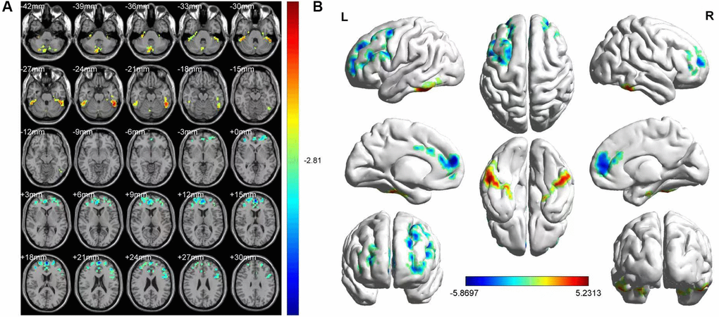
Figure 2.FMRI showed brain regions where the ALFF values' difference was statistically significant between the HR and HC groups. The difference of ALFF value is shown in (A, B) shows the ALFF changes in the cerebral cortex. Different is shown in the left medial SFG, left MFG, left ICL, vermis, left ITG and right SCL. The yellow and red areas represent an increase in ALFF values; the blue regions reduce ALFF values.