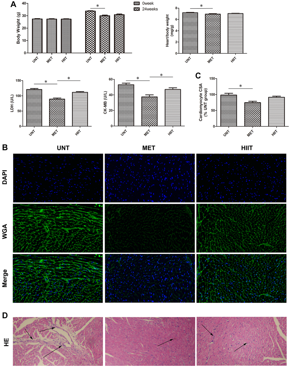
Figure 2.Metabolic data and cardiac tissue damage in the different groups after exercise training. (A) Quantitative analysis of body weight (0 week and 24 weeks), heart/body weight, LDH, and CK-MB levels in the different groups. n = 8 per group. *P < 0.05. (B) Representative photomicrographs of myocardium stained with WGA (green fluorescence) and DAPI (blue fluorescence). (C) Graph showing the cardiomyocyte CSA. (D) H and E staining showing structure damage in cardiac tissue. Magnification 40×. The arrows indicate positively stained cells. n = 3 per group. *P < 0.05.