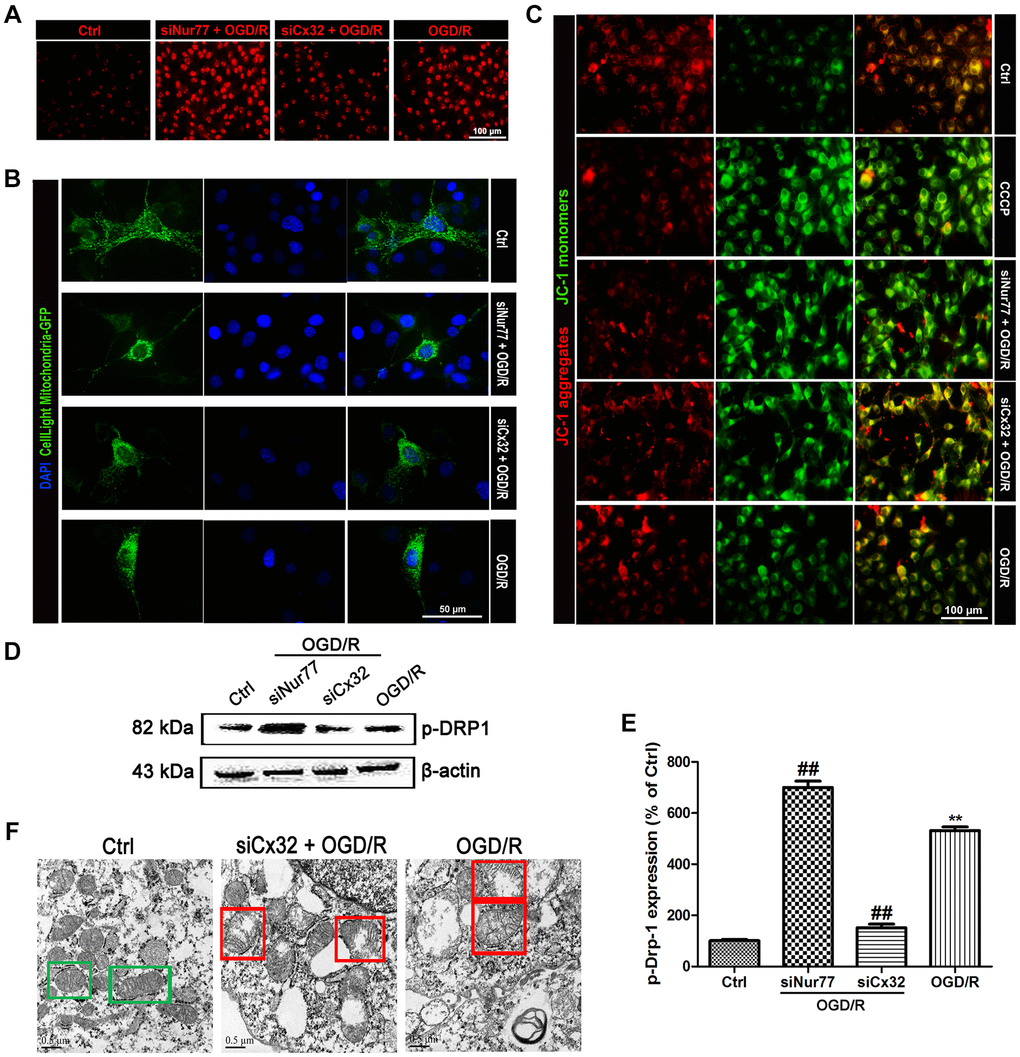
Figure 3.Nur77 attenuated mitochondrial dysfunction after the inhibition of Cx32 following OGD/R injury. Cells were transfected with Cx32 siRNA or Nur77 siRNA for 24 h followed by OGD/R (2 h OGD following by 24 h recovery) before harvested. (A) The level of ROS was detected by DCF-DA. Scale bars, 100 μm. (B) Mitochondria was labeled with GFP (green); cell nuclei were counterstained with DAPI (blue). Photomicrographs were captured under a Nikon Ni-U fluorescence microscope. Scale bars, 50 μm. (C) Δψm was detected by JC-1 dye. Images were shown as the ratio of JC-1 aggregates to JC-1 monomers. Scale bars, 50 μm. (D–E) Representative bands of p-Drp-1, COX4 and TOMM20 protein after OGD/R. Variation in protein loading was determined by blotting with an anti-β-actin antibody. Densitometric scanning of band intensities were calculated as means ± SD (n = 3). **p < 0.01 vs. Ctrl group, ##p < 0.01 vs. OGD/R. (F) Images were collected by transmission electron microscope. Green square represents normal mitochondria with cylindrical shape. Their cristae was well-defined, the double membranes was intact and the density was homogenous; Red square represents abnormal mitochondria. Their cristae was disordered, double membrane was discontinuous, electron density was decreased and minor axis was increased consistent with mitochondrial swelling. Scale bars, 0.5 μm.