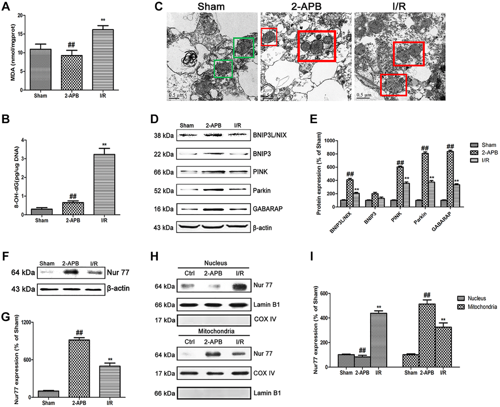
Figure 7.2-APB activated mitophagy through promoting Nur77 translocation from nucleus to mitochondrial after I/R injury. (A) and (B) The levels of MDA and 8-OH-dG were examined at 72 h after reperfusion. (C) Images acquired by transmission electron microscope. Green square represents normal mitochondria with cylindrical shape. Their cristae was well-defined, the double membranes was intact and the density was homogenous; Red square represents abnormal mitochondria. Their cristae was disordered, double membrane was discontinuous, electron density was decreased and minor axis was increased consistent with mitochondrial swelling. Scale bars, 0.5 μm. (D–E) Representative bands of mitophagy-related proteins in brain tissues after I/R. (F–G) Representative bands of Nur77 protein in brain tissues after I/R. (H–I) Mitochondrial translocation of Nur77 was examined by Western blot after I/R. Mitochondria Nur77 expression was normalized against COX IV. Nucleus Nur77 expression was normalized against Lamin B1 expression. Variation in protein loading was determined by blotting with an anti-β-actin antibody. Densitometric scanning of band intensities were calculated as means ± SD (n = 3). **p < 0.01 vs. Sham group, ##p < 0.01 vs. I/R.