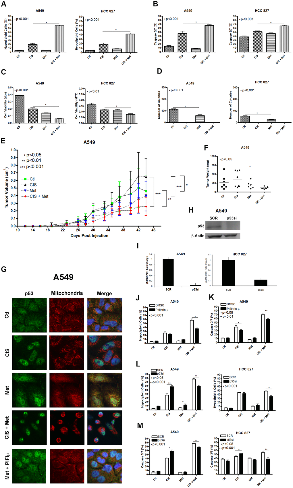
Figure 1.Metformin improves cisplatin-induced death in A549 and HCC 827 NSCLC cells in a P53 dependent manner. Combined treatment between cisplatin and metformin improves DNA fragmentation (p<0.001) (A), caspase 3 and 7 activation (p<0.001) (B) and cell viability assay through MTT (p<0.001 and p<0.01 respectively) (C), when compared to both treatments alone in A549 and HCC 827 cells. Metformin decreased the number of colonies and no colonies was observed after cisplatin treatment in A549 and HCC 827 cells (p<0.001 and p<0.01, respectively) (D). A549 cells injected in NOD/SCID mice also have a smaller volume (p<0.001) (E) and weight (p<0.05) (F) after combined treatment between cisplatin and metformin. Data represent the mean of three independent experiments. Metformin treatment translocate P53 to the mitochondria in A549 cells and this translocation is blocked by pifithrin-μ (G). P53 inhibition by pifithrin-μ protects A549 cells to the metformin induced chemosensitization to cisplatin by decreasing DNA fragmentation (p<0.001) (J) and caspase 3 and 7 activation (p<0.01) (K). TP53 inhibition by siRNA (H, I) also protects A549 and HCC 827 cell from metformin-induced chemosensitization to cisplatin by decreasing DNA fragmentation (p<0.001) (L) and caspase 3 and 7 activation (p<0.001) (M). Data represent the mean of three independent experiments. A549 cells were treated with 10mM of metformin for 72 h and 25μM of cisplatin (with or without metformin) for another 72 h. HCC 827 cells were treated with 20mM of metformin for 72 h and 20μM of cisplatin (with or without metformin) for another 72 h.