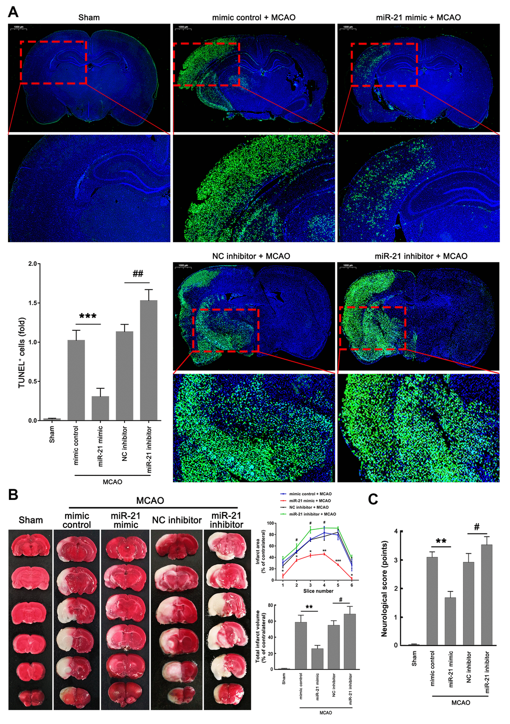
Figure 6.MiR-21/p53/Bcl-2/Bax signaling regulates ischemic neuronal injury in vivo. (A) Representative images showed the TUNEL-labeled cells in the brain slices. Bar graph summarized the numbers of TUNEL+ cells (fold) in the ischemic region. Scale bar, 1000μm. (B) Brain infarction was visualized by TTC staining at 24 h after operation. Curve lines summarized infarct areas in the ipsilateral hemisphere normalized to the total areas of the contralateral hemisphere in sequential coronal brain slices. Bar graph showed the volumes of total cerebral infarct in the ipsilateral hemisphere normalized to the total volumes of the contralateral hemisphere. (C) Neurologic deficits were analyzed by a neurologic deficit score. Eight mice were randomly selected from each treatment group. *, mimic control + MCAO group vs. miR-21 mimic + MCAO group. *P≤0.05, **P≤0.01, ***P≤0.001. #, NC inhibitor + MCAO group vs. miR-21 inhibitor + MCAO group. #P≤0.05, ##P≤0.01.