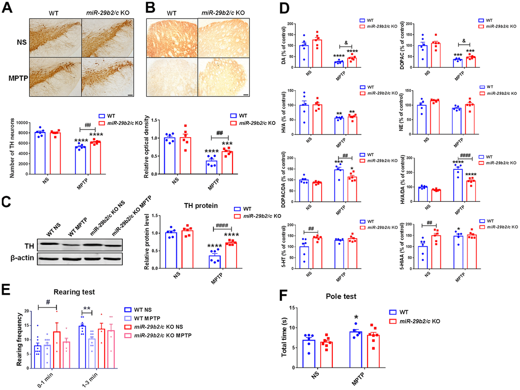
Figure 3.The analysis of the nigrostriatal pathway and behavioral performance of WT and miR-29b2/c KO mice at 3 days after MPTP administration. (A) Immunohistochemical staining of TH in the SNpc of WT and miR-29b2/c KO mice. Scale bar: 0.1mm. Stereological counting of TH positive dopaminergic neurons is shown in the lower panel. n=5-6. (B) Immunohistochemical staining showing striatal TH positive nerve fibers of WT and miR-29b2/c KO mice. Scale bar: 0.05mm. Densitometric analysis of the relative optical density of the staining is shown in the lower panel. n=6. (C) Western blot showing TH protein levels in the striatum of WT and miR-29b2/c KO mice. β-actin served as a loading control. The quantification of the relative TH protein levels is shown in the right panel. n=6. (D) Levels of striatal dopamine (DA), 5-HT, their metabolites, and norepinephrine (NE) in WT and miR-29b2/c KO mice. n=5-6. (E) The rearing frequency of WT and miR-29b2/c KO mice between 0-1min and 1-3min in the Rearing test. n=4-11. (F) The total time of WT and miR-29b2/c KO mice in the Pole test. n=5-7. The differences were analyzed by two-way ANOVA followed by LSD multiple comparison tests. *p<0.05, **p < 0.01, ***p<0.001 and ****p<0.0001, vs normal saline control. # p < 0.05, ## p < 0.01 and ####p<0.0001, vs WT group. & p < 0.05, by Student-T-test.