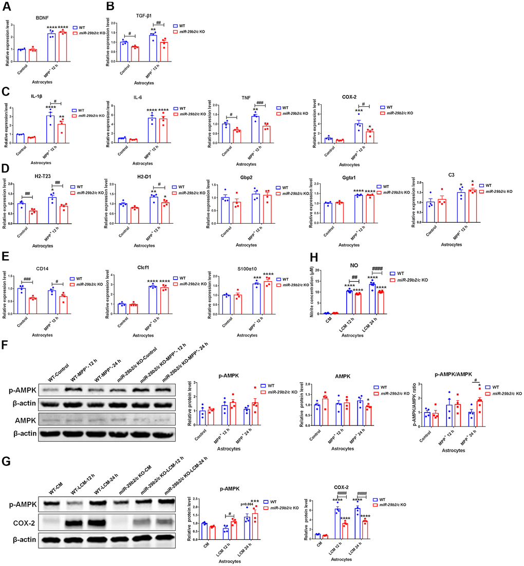
Figure 5.The effects of miR-29b2/c deficiency in MPP+- and conditioned medium-treated primary astrocytes. (A–E) qPCR analysis of BDNF (A), TGF-β1 (B), IL-1β, IL-6, TNF and COX2 (C), H2-T23, H2-D1, Gbp2, Gpta1 and C3 (D), CD14, Clcf1 and S100α10 (E) transcripts in WT and miR-29b2/c KO primary astrocytes treated with PBS or MPP+ for 12 h. n=4. (F) Western blot analysis of p-AMPK and AMPK protein expression in WT and miR-29b2/c KO primary astrocytes treated with PBS or MPP+ for 12 h and 24 h. β-actin served as a loading control. Quantifications of relative p-AMPK and AMPK protein levels and their ratio are shown in the right panel. n=4-5. (G) Western blot analysis of p-AMPK and COX-2 protein expression in WT and miR-29b2/c KO primary astrocytes exposed to conditioned medium (CM) or LPS-treated conditioned medium (LCM) of BV2 cells for 12 h and 24 h. β-actin served as a loading control. Quantifications of relative p-AMPK and COX-2 protein levels are shown in the right panel. n=3-4. (H) Nitrite concentration in the culture medium of WT and miR-29b2/c KO primary astrocytes treated with CM or LCM of BV2 cells for 12 h and 24 h. The differences were analyzed by two-way ANOVA followed by LSD multiple comparison tests. *p<0.05, **p < 0.01, ***p<0.001 and ****p<0.0001, vs PBS control. # p < 0.05, ## p < 0.01, ### p<0.001 and #### p<0.0001, vs WT group.